DermNet provides Google Translate, a free machine translation service. Note that this may not provide an exact translation in all languages
Home Dermatological emergencies Erythema multiforme CME
Dermatological emergencies
Erythema multiforme
Created 2008.
Learning objectives
- Diagnose, classify and manage erythema multiforme
Introduction
Erythema multiforme (EM) is conventionally separated into EM minor and EM major. It is now separated from Stevens Johnson syndrome (SJS) and toxic epidermal necrolysis (T.E.N.).
Clinical features
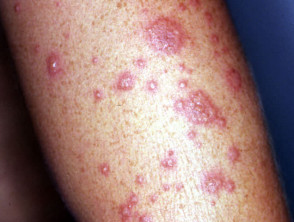
EM minor
EM is more common in men than women and 50% are under 20 years of age. It is an eruption of classic target lesions on the extremities associated with mild fever and malaise. It persists for one to three weeks.
EM minor is mostly preceded by infection. Common causes are:
- Herpes simplex (often labial)
- Orf
- Other viral infections
- Vaccination (diphtheria, tetanus, smallpox)
Drugs are an uncommon cause. Recurrent EM is nearly always due to recurrent herpes simplex.
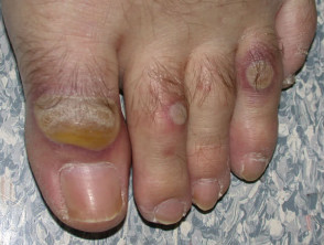
Erythema multiforme minor
EM major
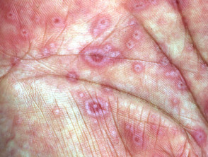
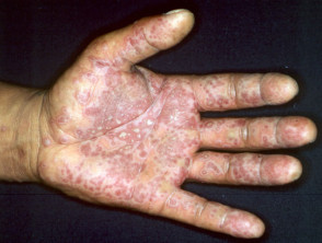
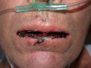
EM major is rare, except in patients suffering from human immunodeficiency virus infection. It is predominantly a mucosal eruption of erosions and blisters in the oropharynx, on the lips, conjunctivae and genitalia accompanied by fever and prostration. Target lesions or acral bullae may also be present.
Like toxic epidermal necrolysis, EM major is usually a drug eruption.
| The most common drugs causing EM major |
|---|
|
Infections are less common causes, but EM major may occur in epidemics associated with Mycoplasma pneumoniae. There is usually lymphopaenia.
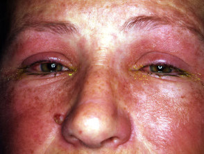
Erythema multiforme major (Stevens-Johnson syndrome)
Investigations
Look for underlying causes and complications of the disease.
- Skin swabs for herpes simplex and bacterial culture
- Full blood count
- Viral titres especially mycoplasma
Skin biopsy findings are often diagnostic:
- Necrotic keratinocytes (especially EM major)
- Spongiosis or intraepidermal vesicles
- Basal layer liquefaction and/or subepidermal blister
- Mixed perivascular inflammatory infiltrate
- Red blood cell extravasation
Management
EM minor resolves in 10 days or so. Symptomatic treatment may include:
- Incise and drain large bullae (leave the blister intact if possible)
- Topical corticosteroids
- Oral antihistamines
Recurrent EM minor can be minimised or prevented by prophylactic oral acyclovir.
Most cases of EM major require hospitalisation for supportive care. This may include:
- Intravenous fluid replacement
- Mouth care (antiseptic and analgesic mouth washes)
Oral corticosteroids should be avoided. In severe cases, EM major should be managed in an Intensive Care facility as for toxic epidermal necrolysis.
Activity
What other skin diseases may cause target lesions?