DermNet provides Google Translate, a free machine translation service. Note that this may not provide an exact translation in all languages

Translate

Progressive macular hypomelanosis

Author: Dr Lydia Chan, Medical Registrar, Waikato Hospital, Hamilton. Chief Editor: Dr Amanda Oakley, Dermatologist, Hamilton, New Zealand, December 2014.


toc-icon

What is progressive macular hypomelanosis?

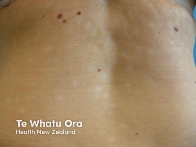
Progressive macular hypomelanosis is a common skin condition presenting as circular areas of pale skin (hypopigmentation) mostly located on the trunk.

Who gets progressive macular hypomelanosis?

Progressive macular hypomelanosis affects teenagers and adults worldwide. It is more common in people of darker skin colour that light skin colour, and may be more common in women than in men.

What are the clinical features of progressive macular hypomelanosis?

Progressive macular hypomelanosis mainly affects the trunk, rarely extending to the arms, legs and/or neck. There are multiple, circular, poorly defined pale and non-scaly spots that merge together.

The patches are not scaly, unlike hypopigmented pityriasis versicolor, which can also cause pale patches on the trunk. They are not preceded by inflammation, unlike postinflammatory hypopigmentation following nummular eczema or psoriasis.

The course of progressive macular hypomelanosis is reported to be variable. Some cases may resolve in 3–5 years. Others remain stable and yet others progress over decades and may then resolve in middle age.

Progressive macular hypomelanosis

What is the cause of progressive macular hypomelanosis?

The cause of progressive macular hypomelanosis is not known. The acne bacteriaCutibacterium acnes, have been found in hair follicles of hypopigmented areas and may cause the loss of colour. However, these bacteria have not been found in all patients with progressive macular hypomelanosis. Genetic factors are also thought to be important.

How is the diagnosis of progressive macular hypomelanosis made?

Progressive macular hypomelanosis is usually diagnosed by clinical suspicion and supported by the presence of fluorescence on Wood light examination. The hair follicles fluoresce with a red glow, indicating they are colonised by P. acnes.

Dermoscopy shows poorly-defined white macules with faint reticular pigmentation. The macules are usually not scaly but focal scale is occasionally seen.

Microscopic examination of a skin biopsy shows a reduction in melanin in the the affected skin, with normal numbers of melanocytes (specialised cells that produce melanin). On electron microscopy of affected areas, the melanosomes (the granules that store melanin in the cell) are smaller and aggregated, compared to surrounding skin.

How is progressive macular hypomelanosis treated?

Treatments for progressive macular hypomelanosis include:

Isotretinoin was effective in one case report.

The condition may relapse following treatment.


References

  • Relyveld GN, Menke HE, Westerhof W. Am J Clin Dermatol. 2007;8(1):13–9. Progressive macular hypomelanosis: an overview. PubMed
  • Perman M, Sheth P, Lucky AW. Pediatr Dermatol 2008 Jan-Feb; Vol. 25 (1), pp. 63–5. Progressive macular hypomelanosis in a 16-year old. PubMed
  • Relyveld G, Menke H, Westerhof W. J Eur Acad Dermatol Venereol 2006 Nov; Vol. 20 (10), pp. 1363–4. Progressive and extensive hypomelanosis and extensive pityriasis alba: same disease, different names? PubMed
  • Relyveld GN, Dingemans KP, Menke HE, Bos JD, Westerhof W. Ultrastructural findings in progressive macular hypomelanosis indicate decreased melanin production. J Eur Acad Dermatol Venereol. 2008 May;22(5):568–74. PubMed
  • Kwah YC, Chong WS, Thiam-Seng Theng C, Goh BK. Photodermatol Photoimmunol Photomed. 2010;26(3):153-5. Treatment of Progressive macular hypomelanosis with narrow-band ultraviolet B phototherapy. PubMed
  • Kuznetsov AV, Flaig MJ, Ruzicka T, Herzinger T. J Clin Pathol 2011;(8):734-6. Progressive macular hypomelanosis Guillet-Hélénon: structural and immunohistochemical findings. PubMed
  • Sim JH, Lee DJ, Lee JS, Kim YC. Comparison of the clinical efficacy of NBUVB and NBUVB with benzoyl peroxide/clindamycin in progressive macular hypomelanosis. J Eur Acad Dermatol Venereol. 2011 Nov;25(11):1318–23. PubMed
  • Kim YJ, Lee DY, Lee JY. J Dermatol. 2012;39(11):937–38. Progressive macular hypomelanosis showing excellent response to oral isotretinoin. PubMed
  • Errichetti E, Stinco G. Dermoscopy in general dermatology: a practical overview. Dermatol Ther (Heidelb). 2016;6(4):471–507. doi:10.1007/s13555-016-0141-6. PubMed

On DermNet

Other websites

Books about skin diseases


Related information

Sign up to the newsletter