DermNet provides Google Translate, a free machine translation service. Note that this may not provide an exact translation in all languages

Translate

Principles of dermatological practice

Structure of the dermis and subcutis

Created 2008.

Learning objectives

  • Name specific structural components of the basement membrane zone, dermis and subcutis

Introduction

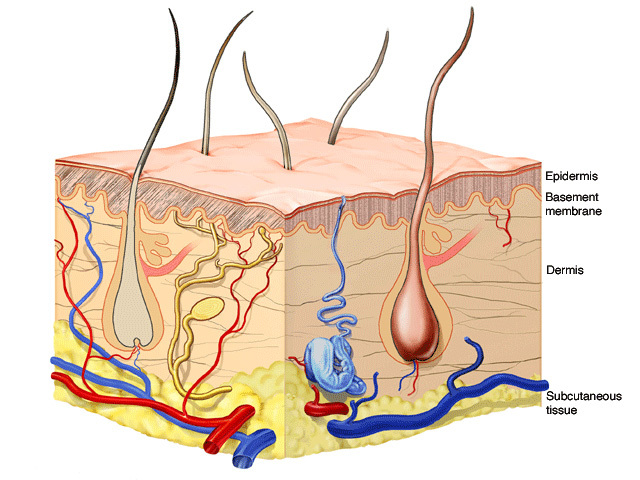
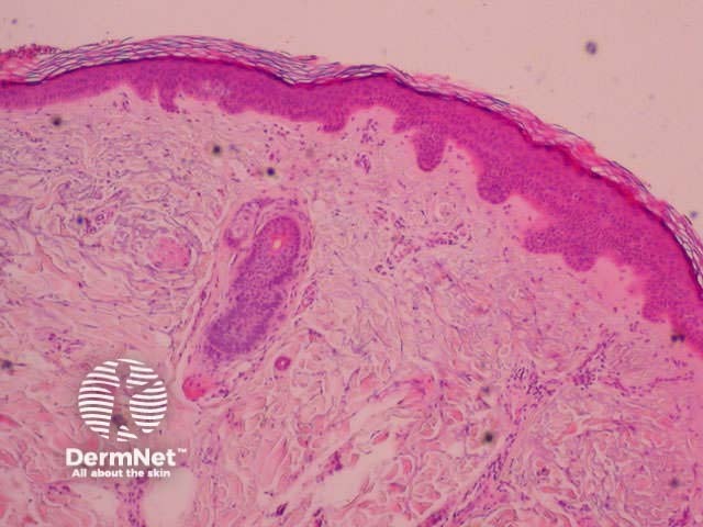
The basement membrane zone is the communication channel between epidermis and dermis. The dermis supports the epidermis, providing nutrients and protecting it.

The dermis supports the epidermis by providing it with nutrients and toughness. It is made up of fibres and ground substance, with nerves, blood vessels and cellular infiltrations. The papillary dermis is the upper portion beneath the epidermis, characterised by thin haphazardly arranged collagen fibres, thin elastic fibres and ground substance. The lower portion is the reticular dermis, composed of coarse elastic fibres and thick collagen bundles parallel to the skin surface.

The dermis is full of double rows of peg-like formations called papillae under the basement membrane zone. Each double row underlies an epidermal ridge.

The papillary dermis is the portion of the dermis just below the epidermis. The reticular dermis extends from the papillary dermis to the fat.

Below this is subcutaneous tissue, the shock absorbing, and insulating and energy storage layer.

Normal skin

Basement membrane zone

StructureDescription
Basal cell membrane
  • Selectively permeable membrane.
  • Hemidesmosomes on dermal side.
Lamina lucida
  • Although appears clear under electron microscope, it is a dense structure holding anchoring filaments (type 7 collagen), fibronectin and laminin (a glycoprotein).
  • Contains pemphigoid antigens.
Lamina densa
  • Appears dense under electron microscope.
  • Made of type 4 collagen and heparan sulphate.
  • Contains epidermolysis bullosa acquisita antigen.
Sublamina densa
  • Cross banded fibrils bind lamina densa to papillary collagen.

Dermis

StructureDescription
Collagen
  • The major protein of the dermis and 90% of the dermal fibres.
  • Provides tensile strength.
  • Fibres are cross-linked triple helix of polypeptide chains.
  • Continuously synthesised by fibroblasts and degraded by collagenase.
  • Haphazard arrangement in papillary dermis; bundles are parallel to surface in reticular dermis.
  • Fine network of Type 3 collagen around blood vessels stains with silver.
Elastic fibres
  • Comprise 10% of the fibres in the dermis.
  • Return deformed skin to its resting state.
  • Made of amorphous elastin protein, surrounded by microfibrils.
  • Synthesised by fibroblasts.
  • Thin fibres in papillary dermis; thicker in reticular dermis.
Ground substance
  • Amorphous viscoelastic gel made of anionic polysaccharides (glycosaminoglycans): hyaluronic acid, dermatan sulphate, and chondroitin-6-sulphate.
  • Produced and degraded by fibroblasts and mast cells.
  • Binds water (up to 1000 times own weight); controls flow of solutes.
Fibroblasts
  • Derived from mesenchyme.
  • Produce collagen, elastin, ground substance and fibronectin (a glycoprotein).
Blood vessels
  • Arteries in subcutaneous tissue send arborising branches into the dermis.
  • Lined by endothelial cells; sheathed in collagen; surrounded by pericytes, dendritic macrophages and T-lymphocytes.
  • Hypothalamic control results in constriction and dilatation with heat, emotional stress (blush with embarrassment, anger; blanch with fear) and spicy food.
Lymphatics
  • Extensive network throughout dermis.
Nerves
  • Extensive network of sensory and autonomic nerve fibres.
  • Distinct sensory nerve endings for touch, heat, cold, pressure, pain.
Arrector pili muscles
  • Originate near the basement membrane zone and attach to the hair follicle near its base.
  • Smooth muscles.
  • Cause erection of the hairs on exposure to cold or fear (goose bumps).
  • Best developed on areolae and tunica dartos of scrotum.
Immune cells
  • Lymphocytes, neutrophils, monocytes (histiocytes and activated macrophages), mast cells.
  • Most often found around blood vessels.

Subcutaneous tissue (subcutis, hypodermis)

StructureDescription
Adipose cells
  • Connective tissue (lipocytes).
  • Groups of cells form lobules with wide variations.
  • Surrounded by connective tissue, larger blood vessels and nerves.
  • For fat storage, heat insulation, shock absorption.

Activity

List collagen subtypes, their differences, location and function.


Related information

References:

Other websites:

Books about skin diseases:

See the DermNet NZ bookstore

Sign up to the newsletter