DermNet provides Google Translate, a free machine translation service. Note that this may not provide an exact translation in all languages

Translate

Majocchi granuloma

Author: Dr Maneka Gnanasegaram, Dermatology Registrar, Greenlane Hospital, Auckland, New Zealand, 2012. Updated by Dr Jannet Gomez. DermNet NZ Editor in Chief: Adjunct Assoc. Prof. Amanda Oakley, Hamilton, New Zealand. December 2018.


toc-icon

What is Majocchi granuloma?

Majocchi granuloma is a deep and persistent suppurative and granulomatous folliculitis (hair follicle infection) caused by fungal infection. Majocchi granuloma is also known as granuloma trichophyticum.

Who gets Majocchi granuloma?

Various forms of tinea are common in males and females worldwide. Majocchi granuloma mostly affects adults. It is common in developing countries. The associated factors leading to deep follicular infection by dermatophyte fungi may include:

What is the cause of Majocchi granuloma?

Majocchi granuloma is due to disruption of infected hair follicles so that hair shafts and fungi penetrate into the dermis and subcutaneous tissue. 

The infection is most often caused by Trichophyton rubrum. Other fungi that may cause Majocchi granuloma include:

  • T. mentagrophytes
  • T. tonsurans
  • T. violaceum
  • Epidermophyton floccosum
  • Non-dermatophyte moulds, like Aspergillus and Phoma.

What are the clinical features of Majocchi granuloma?

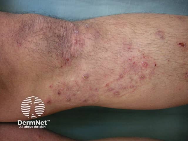
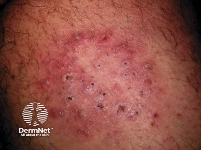
Majocchi granuloma presents with an irregular red, scaly plaque in which there are follicular papules, pustules and nodules. It is usually found on one lower leg.

Evidence for fungal infection may be found in other sites such as scaling on the sole of the foot (tinea pedis) or yellowed, irregular toenails (tinea unguium).

There are two forms of Majocchi granuloma.

  • The perifollicular papular form is a localised dermal infection and usually occurs in healthy individuals
  • The deep subcutaneous plaque or nodular form arises in people with underlying immunosuppression.

Majocchi granuloma

How is Majocchi granuloma diagnosed?

Suspicion is raised due to the clinical appearance of Majocchi granuloma.

To confirm a fungal infection, scrapings and hair samples may be taken from the affected area for microscopy and fungal culture (mycology). Granulomatous inflammation is found on skin biopsy.

What is the treatment for Majocchi granuloma?

The recommended treatment for Majocchi granuloma is a 4  to 6-week course of oral antifungal agent.

Topical antifungal agents may not be effective due to the deep invasion of the fungus into the hair follicle.


References

  • Dermatology. Bolognia JL, Jorizzo JL, Rapini RP. Second edition. Mosby Elsevier
  • Radentz WH, Yanase DJ. Papular lesions in an immunocompromised patient. Trichophyton rubrum granulomas (Majocchi's granuloma). Arch Dermatol 1993; 129 (9): 1189-90, 1192-3.  PubMed.
  • Tse KC, Yeung CK, Tang S, Chan HH, Li FK, Chan TM, et al. Majocchi's granuloma and posttransplant lymphoproliferative disease in a renal transplant recipient. Am J Kidney Dis. 2001;38: PubMed.
  • Liao YH, Chu SH, Hsiao GH, Chou NK, Wang SS, Chiu HC. Majocchi's granuloma caused by Trichophyton tonsurans in a cardiac transplant recipient. Br J Dermatol. 1999;140:1194–1196. PubMed
  • Kim ST, Baek JW, Kim TK, Lee JW, Roh HJ, Jeon YS, et al. Majocchi's granuloma in a woman with iatrogenic Cushing's syndrome. J Dermatol 2008 Dec;35(12):789-91. doi: 10.1111/j.1346-8138.2008.00571.x. PubMed PMID: 19239561.

On DermNet

Other websites

Books about skin diseases


Related information

Sign up to the newsletter