DermNet provides Google Translate, a free machine translation service. Note that this may not provide an exact translation in all languages

Translate

Malassezia folliculitis

Author: Dr Amanda Oakley, Dermatologist, Hamilton, New Zealand,1997. Updated by Dr Thomas Stewart, General Practitioner, Sydney, Australia, November 2017. Revised September 2020.


toc-icon

What is Malassezia folliculitis?

Malassezia folliculitis, also known as pityrosporum folliculitis, is an infection of the pilosebaceous unit caused by lipophilic Malassezia yeasts particularly M. globosa, M. sympodialis and M. restricta. Malassezia yeast are normal inhabitants of the human skin surface and only cause disease under specific conditions.

Malassezia yeasts have been linked to a number of other skin diseases including seborrhoeic dermatitis and pityriasis versicolor.

Malassezia folliculitis

Who gets Malassezia folliculitis?

Malassezia folliculitis is most commonly seen in adolescent and young adult males.

Risk factors for developing Malassezia folliculitis include:

What are the clinical features of Malassezia folliculitis?

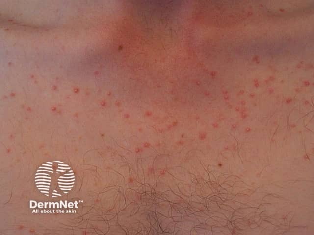
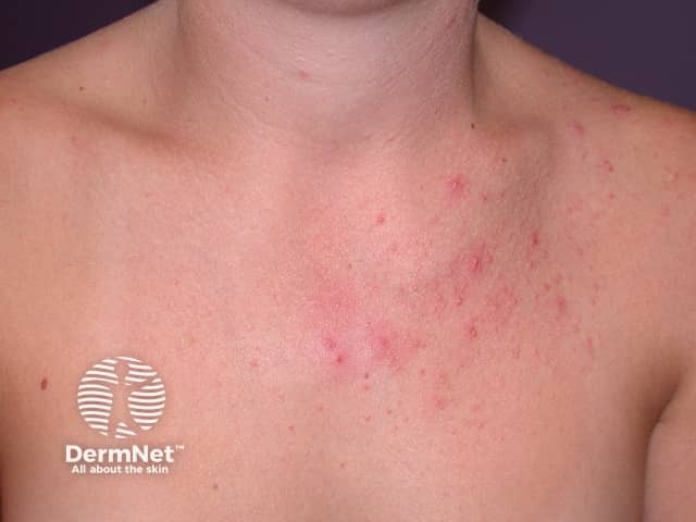
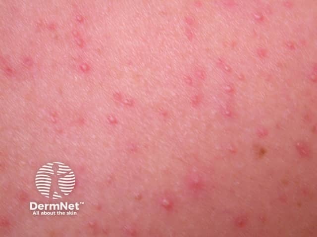
Malassezia folliculitis presents as small, uniform, itchy papules and pustules particularly on the upper back and chest. Other sites involved can include the forehead/hair line, chin, neck, and extensor aspect of the upper limbs. This is a monomorphic eruption and comedones are not seen, distinguishing this condition from acne vulgaris.

Wood lamp examination may demonstrate a yellow-green fluorescence.

Dermoscopy features have been described and include:

  • Papules and pustules based on hair follicles
  • Perifollicular erythema
  • Perilesional scale
  • Hypopigmented and coiled/looped hairs.

How is Malassezia folliculitis diagnosed?

Clinical examination raises the diagnosis. Laboratory investigations on skin scraping, tape stripping, swab, or skin biopsy confirm the presence of yeasts as bacterial folliculitis and steroid acne can be clinically similar.

  • Potassium hydroxide preparation of skin scrapings may reveal budding yeasts. Hyphae are rarely seen.
  • Other stains such as the May-Grünwald-Giema stain are less commonly used.
  • Culture of Malassezia species typically requires special media for growth, so is not routinely performed.
  • Yeast are seen within the hair follicle and possibly the surrounding dermis on histopathological examination. 

What is the differential diagnosis of Malassezia folliculitis?

Diagnosis of Malassezia folliculitis is often delayed due to its clinical resemblance to:

  • Acne vulgaris
  • Steroid acne
  • Bacterial folliculitis.

What is the treatment of Malassezia folliculitis?

It is important to address any predisposing factors at the outset, as Malassezia folliculitis has a tendency to recur.

Topical treatments such as selenium sulfide shampoo, econazole solution and topical ketoconazole are effective in the majority of cases but may require a longer course than systemic agents. Ongoing weekly application may be particularly useful as maintenance therapy to prevent recurrence.

Oral treatment may be more effective than topical, although results of clinical trials have not consistently shown this. Fluconazole is used more commonly than itraconazole due to its superior side effect profile.

Isotretinoin and photodynamic therapy (PDT) have been used with some success in small case series of recalcitrant disease.

How can Malassezia folliculitis be prevented?

Recurrence is common, even after successful treatment.

Long-term prophylaxis with topical agents may be considered in those at high-risk or with multiple recurrences.

Periodic re-evaluation of predisposing factors is recommended. 


Bibliography 

  • Durdu M, Güran M, Ilkit M. Epidemiological characteristics of Malassezia folliculitis and use of the May-Grünwald-Giemsa stain to diagnose the infection. Diagn Microbiol Infect Dis. 2013;76(4):450-7. doi:10.1016/j.diagmicrobio.2013.04.011. PubMed.
  • Akaza N, Akamatsu H, Sasaki Y, et al. Malassezia folliculitis is caused by cutaneous resident Malassezia species. Med Mycol. 2009;47(6):618-24. doi:10.1080/13693780802398026. PubMed.
  • Gaitanis G, Velegraki A, Mayser P, Bassukas ID. Skin diseases associated with Malassezia yeasts: facts and controversies. Clin Dermatol. 2013;31(4):455-63. doi:10.1016/j.clindermatol.2013.01.012. PubMed
  • Rubenstein RM, Malerich SA. Malassezia (pityrosporum) folliculitis. J Clin Aesthet Dermatol. 2014;7(3):37-41. PubMed.
  • Hald M, Arendrup MC, Svejgaard EL, et al. Evidence-based Danish guidelines for the treatment of Malassezia-related skin diseases. Acta Derm Venereol. 2015;95(1):12-19. doi:10.2340/00015555-1825. PubMed.
  • Bäck O, Faergemann J, Hörnqvist R. Pityrosporum folliculitis: a common disease of the young and middle-aged. J Am Acad Dermatol. 1985;12(1 Pt 1):56-61. doi:10.1016/s0190-9622(85)70009-6. PubMed.
  • Prindaville B, Belazarian L, Levin NA, Wiss K. Pityrosporum folliculitis: a retrospective review of 110 cases. J Am Acad Dermatol. 2018;78(3):511-14. doi:10.1016/j.jaad.2017.11.022. PubMed
  • Lee JW, Kim BJ, Kim MN. Photodynamic therapy: new treatment for recalcitrant Malassezia folliculitis. Lasers Surg Med. 2010;42(2):192-6. doi:10.1002/lsm.20857. PubMed.
  • Saunte DML, Gaitanis G, Hay RJ. Malassezia-associated skin diseases, the use of diagnostics and treatment. Front Cell Infect Microbiol. 2020;10:112. doi:10.3389/fcimb.2020.00112. PubMed
  • Jakhar D, Kaur I, Chaudhary R. Dermoscopy of pityrosporum folliculitis. J Am Acad Dermatol. 2019;80(2):e43-4. doi:10.1016/j.jaad.2018.08.057. PubMed

On DermNet

Other websites

Books about skin diseases


Related information

Sign up to the newsletter