Introduction
Demographics
Causes
Spread
Occurrence
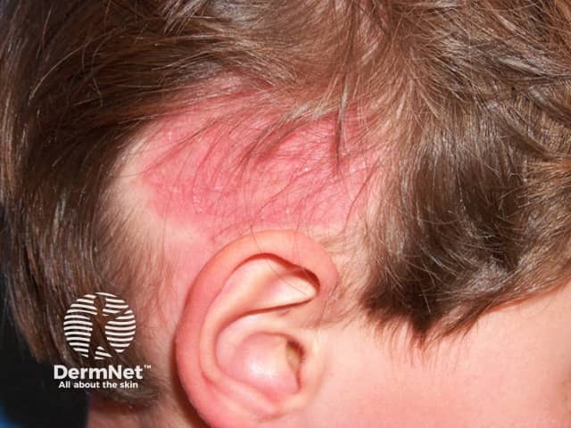
Clinical features
Complications
Diagnosis
Differential diagnoses
Treatment
Outcome
Tinea capitis is a fungal infection of the scalp, involving both the skin and hair. It is also known as scalp ringworm. Symptoms of tinea capitis include hair loss, dry scaly areas, redness, and itch. Tinea barbae is essentially the same condition involving the beard area.


Tinea capitis predominantly affects preadolescent children, with incidence peaking between the ages of three and seven years. It can also affect adults, particularly those who are immunocompromised. Tinea capitis is found in most parts of the world, although the prevalence of a particular fungal species causing tinea capitis varies geographically. Risk factors include animal contact, household crowding, lower socioeconomic status, warm humid environments, and contact sport. The introduction of antifungal agents, population movements, and improved hygiene practices are associated with evolving patterns of infection.
Tinea capitis is caused by dermatophytic fungi capable of invading keratinised tissue, such as the hair and nails. While over 40 different species of dermatophytes are known to exist, only a small number are associated with tinea capitis. Dermatophytes may be classified into three broad categories, according to host preference: anthropophilic species (humans), zoophilic species (animals), and geophilic species (soil).
In New Zealand, Europe, and Asia the most common causative agent is the zoophilic species Microsporum canis which originates in cats.
Examples of other zoophilic fungi that cause tinea capitis include:
Trichophyton tonsurans is an anthropophilic dermatophyte that is the most common cause of tinea capitis in the United States. T. violaceum is an anthropophilic fungus seen in New Zealand in patients who have migrated from Africa or the Middle East.
Examples of other anthropophilic fungi that cause tinea capitis include:
Geophilic fungi, such as M. gypseum, originate in the soil and are rare causes of tinea capitis.
Tinea capitis is a contagious infection. Anthropophilic species are spread following contact with infected persons, including asymptomatic carriers, or contaminated objects (fomites). Fomites that can harbour anthropophilic dermatophytes include hairbrushes, hats, towels, bedding, couches, and toys; fungal spores may remain viable on these for months. Zoophilic species are transmitted from infected animals, including household pets (especially kittens) or stray cats and dogs. Zoophilic species can spread person-to-person.
Following invasion of the keratinised stratum corneum of the scalp (see structure of the normal skin), the fungus grows downwards into the hair follicle and the hair shaft. It penetrates the hair cuticle and typically invades the hair shaft in one of three ways:
Clinical features vary according to the species of dermatophyte, type of hair invasion, and the extent of the inflammatory host response. In all types, partial hair loss with some degree of inflammation is characteristic.
The clinical features may be broadly categorised into non-inflammatory and inflammatory variants.
Non-inflammatory variants of tinea capitis infection include the following.
Inflammatory variants of tinea capitis infection include the following.
The fungal infection may extend to involve other hair-bearing sites including eyebrows and eyelashes, and regional lymphadenopathy may be present.


Alopecia can result in psychosocial distress for the patient, especially when scarring alopecia following inflammatory tinea capitis results in permanent bald patches. A secondary rash may occur with inflammatory tinea capitis, particularly after initiating antifungal treatment; this is known as a dermatophytide or id reaction. Rarely, erythema nodosum has been known to occur. Secondary bacterial infection may develop.
Tinea capitis is usually suspected clinically but should be confirmed on microscopy and culture (see laboratory tests for fungal infections).
Wood lamp examination is diagnostic when hair fluorescence is seen, although few fungi cause infected hairs to fluoresce. Bright green fluorescence of infected hairs is observed in tinea capitis caused by Microsporum species (M. ferruginium, audouinii, canis, and distorum). Identifying affected hairs in this way may help with obtaining an appropriate specimen for microscopy and culture. Wood lamp examination is of no value in nonfluorescent endothrix Trichophyton infection, with the exception of T. schoenleinii, which can fluoresce a dull grey-green.

Dermoscopy of the scalp (trichoscopy) is a fast and non-invasive procedure useful for confirming tinea capitis so that treatment can be started while waiting for culture results.
Dermoscopic findings characteristic of tinea capitis with a high predictive value but not seen in every case include:
Corkscrew hairs are typical of Trichophyton infection and are seen less commonly in infection due to M. canis.
Signs seen commonly on dermoscopy of tinea capitis, but which are not diagnostic include:


Microscopy of scalp scrapings or plucked hairs can rapidly confirm the diagnosis of tinea capitis. Specimens are wet-mounted in potassium hydroxide and examined under a light microscope for the presence of hyphae and spores.
Fungal culture allows identification of the causative agent, and hence the possible source of infection. However, it is slow, taking up to 4 weeks, and treatment is usually required before the result is available.


Polymerase chain reaction (PCR) screening may result in faster and more sensitive ways of detecting dermatophyte infection, but is not yet widely available.
Sometimes the diagnosis of tinea capitis is made on skin biopsy when characteristic changes are seen (see tinea capitis pathology).
The differential diagnosis list is extensive and includes any condition which may present with patchy alopecia, inflammation, or scaling of the scalp. Examples include:
Tinea capitis always requires at least 4 weeks of systemic medication, as topical agents cannot penetrate the root of the hair follicle. Griseofulvin has previously been the most widely used medication to treat tinea capitis, but it is no longer available in some countries, including New Zealand. Newer antifungal agents such as terbinafine, itraconazole, and fluconazole are at least as effective as griseofulvin for trichophyton infections but less effective for microsporum species.
Topical agents such as povidone-iodine, ketoconazole, and selenium sulfide shampoos can be used to reduce spore transmission.
All members of the household should be screened for tinea capitis and treated simultaneously if found to be affected. Sharing of potential fomites such as hairbrushes, hats, and pillows should be discouraged, and these should be properly cleaned.
In cases of zoophilic infection, family pets should be checked by a veterinarian and treated accordingly.
Non-inflammatory tinea capitis carries an excellent prognosis with appropriate and early treatment. Severe inflammatory tinea capitis may result in areas of permanent alopecia.

