DermNet provides Google Translate, a free machine translation service. Note that this may not provide an exact translation in all languages

Translate

Cutaneous horn

Author: Dr Tina Tian, Medical Oncology Registrar, Hawke's Bay Hospital, Hastings, New Zealand, 2013.


toc-icon

What is a cutaneous horn?

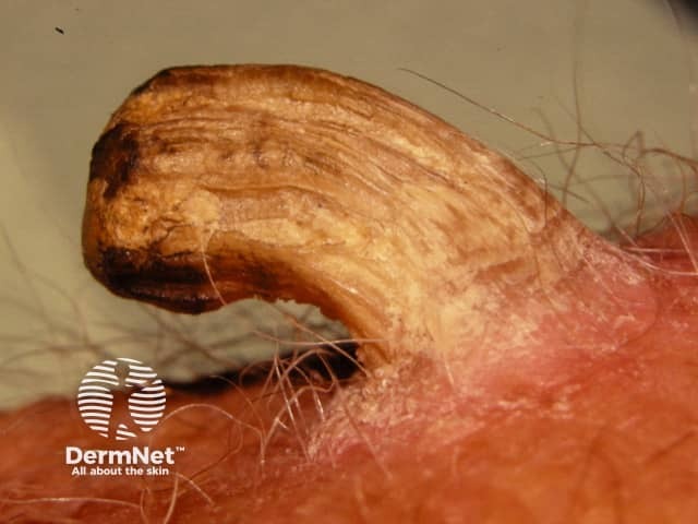
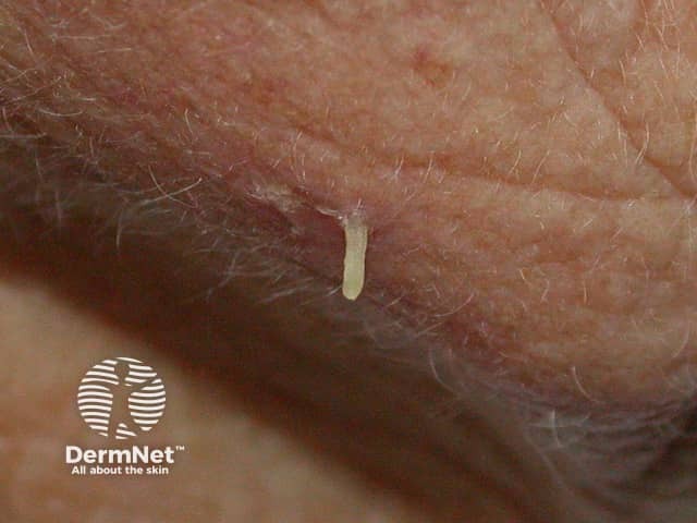
A cutaneous horn (cornu cutaneum) is a hard conical projection from the skin, made of compact keratin. They are so named as they resemble an animal’s horn. They arise from benign, premalignant or malignant skin lesions.

Cutaneous horns

See more images of cutaneous horns ...

Who gets a cutaneous horn?

  • A cutaneous horn is more common in older patients, with the peak incidence in those between 60 and 70.
  • They are equally common in males and females, although there is a higher risk of the lesion being malignant in men.
  • They are more common in people with fairer skins (skin phototype I and 2).

What causes a cutaneous horn?

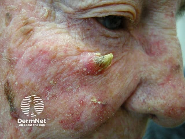
Around half of horns have a benign base, and half are premalignant or malignant. The most common underlying lesions are seborrhoeic keratosis, viral warts (due to human papillomavirus), actinic keratosis, and well-differentiated squamous cell carcinoma (associated with exposure to the sun and other sources of UV radiation).

What are the clinical features of a cutaneous horn?

Appearance

  • A cutaneous horn generally presents as a straight or curved, hard, yellow-brown projection from the skin.
  • It can be surrounded by normal skin or have a border of thickened skin.
  • The side of the horn may be terrace-like or oyster shell-like with horizontal ridges.
  • The base of the horn may be flat, protruding, or like a crater.
  • Inflammation may be present, due to recurrent injury.

Size

  • Typically, the horn is taller than twice the width at the base.
  • It may vary from a few millimetres to several centimetres in size.
  • Giant horns exist — the largest described is a 76-year-old Parisian woman named Madame Dimanche (Widow Sunday) in the 19th century, who grew a horn from her forehead that was 25-cm long.

Location

  • Cutaneous horns are usually singular but can be multiple.
  • They can occur anywhere on the body, but are more common on sun-exposed areas especially the head and ears, back of hands and forearms.
  • They may also occur on the chest, neck, shoulder and penis.

Symptoms

Cutaneous horns are usually asymptomatic. They can be injured causing pain and inflammation.

Worrying features suggestive of malignancy

Whilst no certain features can confidently confirm or exclude malignant lesions, malignant lesions are more common in older patients and in males compared to females. Squamous cell carcinoma is also likely if the horn has the following features:

  • Painful
  • Large size
  • Induration at the base
  • The anatomic site on the nose, ears, backs of hands, scalp, forearms, face and penis
  • Wide base or low height to base ratio
  • Redness at the base of the horn base
  • Lack of terrace formation, due to rapid unorganised growth.

Diagnosis and investigation of cutaneous horn

  • A cutaneous horn is diagnosed by its clinical appearance.
  • Histological examination of the horn base is crucial to rule out malignancy, as there are no certain clinical features that can definitively distinguish benign lesions from skin cancer.
  • The lesion is usually completely excised. In some cases, a deep partial biopsy is taken to establish the diagnosis.

On histology, there is a thickening of the stratum corneum or hyperkeratosis. Orderly horizontal parallel layers of keratin are associated more with benign lesions. Rapidly growing malignant lesions exhibit more erratic growth. Acanthosis is often noted. The base of the lesion shows features of the underlying lesion.

Skin conditions associated with cutaneous horns
Benign Premalignant or malignant

How is a cutaneous horn treated?

Cutaneous horns are usually excised with appropriate margins, dependent of the nature of the lesion.


References

  • Mantese SA, Diogo PM, Rocha A, Berbert AL, Ferreira AK, Ferreira TC. Cutaneous horn: a retrospective histopathological study of 222 cases. An Bras Dermatol. 2010 Mar-Apr 2010;85(2):157–163. PubMed
  • Yu RC, Pryce DW, Macfarlane AW, Stewart TW. A histopathological study of 643 cutaneous horns. Br J Dermatol. May 1991;124(5):449–52. PubMed
  • Pyne J, Sapkota D, Wong JC. Cutaneous horns: clues to invasive squamous cell carcinoma being present in the horn base. Dermatol Pract Concept 2013;3(2):2. PubMed
  • Copcu E, Sivrioglu N, Culhaci N. Cutaneous horns: are these lesions as innocent as they seem to be? World J Surg Oncol. 2004:3(2):18. PubMed
  • Cutaneous Horn — Medscape Reference

On DermNet

Other websites

Text: Miiskin


Related information

Sign up to the newsletter