DermNet provides Google Translate, a free machine translation service. Note that this may not provide an exact translation in all languages

Translate

Desmoplastic melanoma

Author: Hon A/Prof Amanda Oakley, Dermatologist, Hamilton, New Zealand, 2011. Updated by Nigel Maher, Anatomic Pathology Registrar, Sydney, Australia. December 2017.


toc-icon

What is desmoplastic melanoma?

Desmoplastic melanoma is a rare form of invasive melanoma, a skin cancer that arises from pigment cells (melanocytes). 

  • Desmoplastic melanoma often involves nerve fibres, when it is called neurotropic melanoma.
  • The malignant cells within the dermis are surrounded by fibrous tissue.
  • It sometimes arises underneath a lentigo maligna, a type of melanoma in situ.

Desmoplastic melanoma

Management of melanoma is evolving. For up to date recommendations, refer to Australian Cancer Council Clinical practice guidelines for the diagnosis and management of melanoma

Who gets desmoplastic melanoma?

Desmoplastic melanoma accounts for about 1% of melanoma in Australia and New Zealand.

  • It usually occurs in white-skinned individuals.
  • Desmoplastic melanoma is more common in males (65%) than females.
  • Most are older people.

The main risk factors for desmoplastic melanoma are:

  • Increasing age
  • Previous invasive melanoma or melanoma in situ
  • Fair skin that burns easily
  • Sun damaged skin.

What are the clinical features of desmoplastic melanoma?

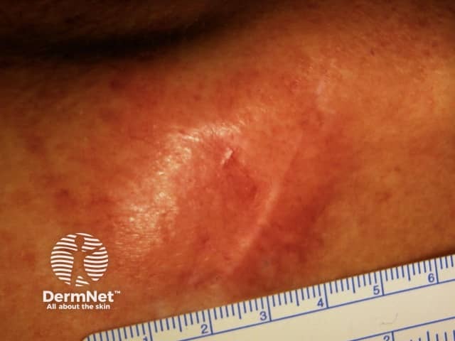
Desmoplastic melanoma is most common on sun-exposed areas of the head and neck (> 50%). Desmoplastic melanoma usually lacks the ABCD melanoma warning signs: Asymmetry, Border irregularity, Colour variation, large Diameter. 

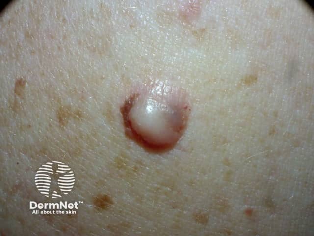
It presents as a slowly enlarging area of thickened skin, sometimes described as scar-like. It is often skin coloured but may be pigmented. ­It becomes more distinctive in time, often growing over months to years before it is recognised. Features include:

  • Firm scar-like consistency with a smooth or irregular surface
  • Flat or raised papule, plaque or nodule  
  • Size  > 6 mm and often 1–2 centimetres in diameter at diagnosis
  • Irregular or regular borders (more often irregular)
  • Variable pigmentation – most often skin-coloured or pink, but may also have areas of brown, grey, blue or black.

Deep invasive melanoma often has the following features:

  • A thickened lesion
  • A greater number of colours, especially blue or black
  • Ulceration or bleeding
  • Itching or stinging.

What is the differential diagnosis of desmoplastic melanoma?

Not uncommonly, desmoplastic melanoma can be mistaken for other benign lesions, such as:

What is the cause of desmoplastic melanoma?

Desmoplastic melanoma is due to the development of malignant pigment cells (melanocytes) within the dermis. These cells may arise within another type of melanoma or in previously normal-appearing skin. What triggers the melanocytes to become malignant is unknown, but it is likely to be a series of changes to DNA.

What tests should be done?

It is essential to diagnose desmoplastic melanoma accurately. A careful clinical history is important, which includes noting previous treatments received for non-resolving lesions. Palpation is an important step, as a large majority of desmoplastic melanoma are indurated. Clinical diagnosis is aided by dermoscopy and skin biopsy (usually excision biopsy). Depending on the thickness and proportion of desmoplasia within the invasive melanoma, sentinel lymph node biopsy, imaging studies and blood tests may be advised.

As desmoplastic melanoma is commonly found on the head and neck, and with lentigo maligna, it is possible that if such lesions are only partially biopsied, the desmoplastic melanoma may be missed.  Reflectance confocal microscopy may be useful to help guide the site of the biopsy.

Dermoscopy

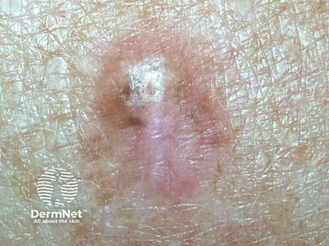
Dermoscopy can be helpful in distinguishing desmoplastic melanoma from other skin lesions.  The most frequently observed dermatoscopic features of desmoplastic melanoma are:

  • Melanocytic features in about 50% (pigmented globules or network)
  • Asymmetrical structure and colours
  • Regression features: scar-like areas, grey dots
  • Multiple colours
  • Atypical or polymorphous vascular pattern
  • Crystalline structures.

Biopsy

If the skin lesion is suspicious of desmoplastic melanoma, it should be urgently cut out (excision biopsy).

The pathological diagnosis of melanoma can be very difficult. Histopathological features of desmoplastic melanoma include:

  • A paucicellular atypical spindle cell proliferation, separated by fibrocollagenous stroma within the dermis or subcutaneous fat.
  • There is overlying melanoma in-situ, typically of lentigo maligna type, in approximately 75% of cases.
  • Nerve infiltration or neurotropism is found in about half of desmoplastic melanomas.
  • Lymphocytic nodular aggregates may be seen in association with desmoplastic melanoma, but are not diagnostic.

Immunohistochemical stains for melanoma may be helpful.

  • S100 and Sox 10 are usually positive.
  • HMB-45 and Melan-A are usually negative.
  • p75 nerve growth factor is a marker for spindle cell melanoma

Pathology report

The pathologist's report should include a macroscopic description of the specimen and melanoma (the naked eye view) and a microscopic description.

  • Diagnosis of primary melanoma
  • Breslow thickness to the nearest 0.1 mm
  • Clark level of invasion
  • Margins of excision, the normal tissue around a tumour
  • Mitotic rate – a measure of how fast the cells are proliferating
  • Whether or not there is ulceration
  • The proportion of the invasive melanoma that is desmoplastic

The report may also include comments about the cell type and its growth pattern, invasion of blood vessels or nerves, inflammatory response, regression and whether there is an associated naevus or another form of melanoma.

Desmoplastic melanoma is categorised into either ‘pure’ or ‘mixed’ subtypes, which reflect the extent of desmoplasia. The mixed subtypes include a component of spindle cell melanoma.

  • Pure subtype: ≥ 90% desmoplasia
  • Mixed subtype: < 90% desmoplasia

What is Breslow thickness?

The Breslow thickness is reported for invasive melanomas. It is measured vertically in millimetres from the top of the granular layer (or base of superficial ulceration) to the deepest point of tumour involvement. In most melanomas, it is a strong predictor of outcome; the thicker the melanoma, the more likely it is to metastasise.

In desmoplastic melanoma, the Breslow thickness is typically thick (3–4 mm). Disease-free survival is worse for melanoma with Breslow thickness > 4 mm, but is slightly more favourable for pure desmoplastic melanoma than other forms of melanoma of the same thickness.  

What is the Clark level of invasion?

The Clark level indicates the anatomic plane of invasion. The deeper the Clark level, the greater the risk of metastasis (secondary spread). It is not considered useful in predicting outcome in desmoplastic melanoma.

What is the treatment for desmoplastic melanoma?

The initial treatment of a desmoplastic melanoma is surgical excision.

  • For pure desmoplastic melanoma, a 2 cm margin of normal tissue may be recommended, whatever its thickness.
  • For mixed desmoplastic melanoma, margins follow guidelines for other forms of invasive melanoma (1–2 cm, depending on Breslow thickness).

Adjuvant radiation therapy may be considered for pure desmoplastic melanoma, Breslow thickness > 4 mm, perineural invasion or positive surgical margins.

Staging

Melanoma staging means analysing primary tumour characteristics (including the Breslow thickness and presence of ulceration), the spread of melanoma to the lymph nodes, the spread of melanoma to loco-regional or distant sites, and the lactate dehydrogenase (LDH) level.

Most melanoma specialists refer to the American Joint Committee on Cancer (AJCC) cutaneous melanoma staging guidelines (8th Edition). The same staging criteria are used in all melanomas, irrespective of their growth pattern.

Stage 0 — In-situ melanoma

Stage 1 — Thin melanoma < 2 mm in thickness

Stage 2 — Thick melanoma > 2 mm in thickness, or > 1mm thickness with ulceration

Stage 3 — Melanoma spread to involve local lymph nodes

Stage 4 — Distant metastases have been detected

Should the lymph nodes be removed?

If the local lymph nodes are enlarged due to metastatic melanoma, they should be completely removed. This requires a surgical procedure, usually under general anaesthetic. If they are not enlarged, they may be tested to see if there is any microscopic spread of melanoma. The test is known as a sentinel node biopsy.

In New Zealand, many surgeons recommend sentinel node biopsy for melanomas thicker than 1 mm, especially in younger persons. However, although the biopsy may help in staging cancer, it does not offer any survival advantage. The necessity for sentinel node biopsy is controversial at present.

  • With pure desmoplastic melanoma, the risk of regional lymph node involvement is reported to be low. Sentinel lymph node biopsy may not be recommended.
  • With mixed desmoplastic melanoma, the risk of regional lymph node involvement is higher, and recommendations are the same as for other types of invasive melanoma.

Lymph nodes containing metastatic melanoma often increase in size quickly. An involved node is usually non-tender and firm to hard in consistency. If this occurs between planned follow-up visits, let your doctor know promptly.

Desmoplastic melanoma has a high mutational load, which may increase the chance of response to immunotherapy with anti-PD-1/PD-1L medications such as pembrolizumab or nivolumab

What happens at follow-up?

The main purpose of follow-up is to detect recurrences early, but it also offers an opportunity to diagnose a new primary melanoma at the first possible opportunity.

The Australian and New Zealand Guidelines for the Management of Melanoma (2008) make the following recommendations for follow-up for patients with invasive melanoma.

  • Self-skin examination
  • Follow-up intervals are preferably six-monthly for five years for patients with stage 1 disease, three-monthly or four-monthly for five years for patients with stage 2 or 3 disease, and yearly after that for all patients.
  • Provide education and support to help the patient adjust to their illness

Follow-up appointments may include:

  • A check of the scar where the primary melanoma was removed: desmoplastic melanoma can recur at the original site (local recurrence)
  • A feel for the regional lymph nodes
  • A general skin examination
  • A full physical examination

In those with more advanced primary disease, follow-up may include:

  • Blood tests, including LDH
  • Imaging: ultrasound, X-ray, CT, MRI and PET scan.

Tests are not typically worthwhile for Stage 1 or Stage 2 melanoma patients unless there are signs or symptoms of disease recurrence or metastasis. No routine tests are necessary for healthy patients who have remained well for five years or longer after removal of their melanoma, whatever stage.

What is the outlook?

The risk of spread and ultimate death from invasive melanoma depends on several factors, but the main one is the measured thickness of the melanoma at the time it was surgically removed. There are conflicting reports about prognostic indicators and outcomes for desmoplastic melanoma, due to its rarity, differences in pathological interpretation, and differing study designs/methodology. The results of the three studies are described below.

  • 5-year overall survival rates were better for 280 patients with localised desmoplastic melanoma (90%, median Breslow thickness 2.5 mm) compared to 7767 patients with non-desmoplastic melanoma (82%, median Breslow thickness 1.6 mm).
  • A study of 1129 desmoplastic melanoma patients in the United States (1992–2007) reported a 5-year specific survival rate of 85% and 10-year survival of 80%. Older age, anatomic site of the head and neck and tumour thickness > 2 mm, ulceration, lymph node involvement and non-receipt of surgery were associated with lower survival.
  • A study of 1,735 desmoplastic melanoma patients, also in the United States (1988–2006), reported overall survival at five years to be 65% and that wide local excision was associated with increased survival. Traditional prognostic factors such as Breslow thickness, nodal positivity, and ulceration did not predict survival in this group of patients.


References

  • Clinical Practice Guidelines for the Management of Melanoma in Australia and New Zealand 2008
  • Richardson et al. The incidence and thickness of cutaneous malignant melanoma in New Zealand 1994–2004. NZ Med J 2008;121:1279. PubMed.
  • Ministry of Health. 2011. Cancer: New registrations and deaths 2008. Wellington: Ministry of Health. Website.
  • Balch CM et al. Final Version of 2009 AJCC Melanoma Staging and Classification. J Clin Oncol 27:6199-6206. PDF file
  • Feng Z, Wu X, Chen V, Velie E, Zhang Z. Incidence and survival of desmoplastic melanoma in the United States, 1992-2007. J Cutan Pathol. 2011 Aug;38(8):616-24.  doi: 10.1111/j.1600-0560.2011.01704.x. Epub 2011 Apr 26. PubMed PMID: 21518379. PubMed
  • Wasif N, Gray RJ, Pockaj BA. Desmoplastic melanoma - the step-child in the melanoma family? J Surg Oncol. 2011 Feb;103(2):158-62. doi: 10.1002/jso.21778. Epub 2010 Dec 22. PubMed
  • Strom T, Caudell J, Han D, et al. Radiotherapy Influences Local Control in Patients With Desmoplastic Melanoma. Cancer. 2014;120(9):1369-78. PubMed
  • Miller D, Emley A, Yang S, et al. Mixed versus pure variants of desmoplastic melanoma: a genetic and immunohistochemical appraisal. Mod Pathol. 2012;25(4):505-15. PubMed
  • Han D, Han G, Zhao X, et al. Clinicopathologic Predictors of Survival in Patients with Desmoplastic Melanoma. PLoS One. 2015;10(3):e0119716. PubMed
  • Maurichi A, Miceli R, Camerini T, et al. Pure desmoplastic melanoma: a melanoma with distinctive clinical behavior. Ann Surg. 2010;252(6):1052-7. PubMed
  • Chen L, Jaimes N, Barker C, et al. Desmoplastic Melanoma: A Review. J Am Acad Dermatol. 2013;68(5):825-33. PubMed
  • Pawlik T, Ross M, Prieto V, et al. Assessment of the role of sentinel lymph node biopsy for primary cutaneous desmoplastic melanoma. Cancer. 2006;106(4):900-6. Journal
  • Quinn M, Crotty K, Thompson J, et al. Desmoplastic and desmoplastic neurotropic melanoma: experience with 280 patients. Cancer. 1998;83(6):1128-35. PubMed
  • George E, McClain S, Slingluff C, et al. Subclassification of desmoplastic melanoma: pure and mixed variants have significantly different capacities for lymph node metastasis. J Cutan Pathol. 2009;36(4):425-32. PubMed
  • Murali R, Shaw H, Lai K, et al. Prognostic factors in cutaneous desmoplastic melanoma: a study of 252 patients. Cancer. 2010;116(17):4130-8. Journal
  • Murali R, Zannino D, Synnott M, et al. Clinical and pathological features of metastases of primary cutaneous desmoplastic melanoma. Histopathology. 2011;58(6):886-95. PubMed
  • de Almeida L, Requena L, Rütten A, et al. Desmoplastic malignant melanoma: a clinicopathologic analysis of 113 cases. Am J Dermatopathol. 2008;30(3):207-15. PubMed
  • Jaimes N, Chen L, Dusza S, et al. Clinical and Dermoscopic Characteristics of Desmoplastic Melanomas. JAMA Dermatol. 2013;149(4):413-21. PubMed
  • Maher N, Solinas A, Scolyer R, et al. Detection of desmoplastic melanoma with dermoscopy and reflectance confocal microscopy. J Eur Acad Dermatol Venereol. 2017;Jun 2. doi: 10.1111/jdv.14381. [Epub ahead of print]. PubMed
  • Shaw H, Quinn M, Scolyer R, et al. Survival in Patients With Desmoplastic Melanoma. J Clin Oncol. 2006;24(8):e12. PubMed

On DermNet

Other websites

Books about skin diseases

Text: Miiskin


Related information

Sign up to the newsletter