Introduction
Demographics
Causes
Clinical features
Diagnosis
Differential diagnoses
Treatment
Outcome
Mastocytoma is the name given to a form of cutaneous mastocytosis in which there is a dermal accumulation of mast cells forming one to thee solitary lesions [1].
Mastocytoma is most often diagnosed in an infant aged 0 to 3 months of age. They are rarely diagnosed in an adult [2]. Mastocytoma is not usually familial.
Mastoma has been associated with a mutation of the KIT gene, which codes for a transmembrane tyrosine receptor on the mast cell responsible for its growth and function [3].
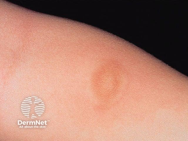
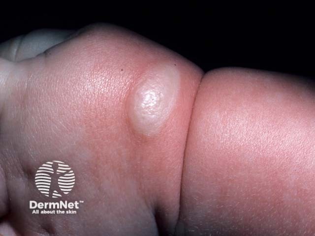
Activation of mast cells causes them to release histamine and other chemicals, which causes localised redness, swelling, itching, and, sometimes, blistering.
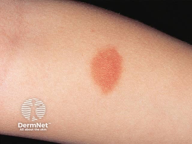
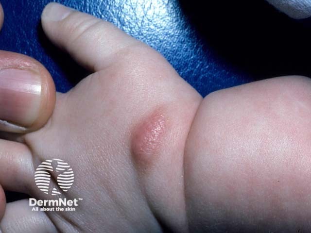
One to three mastocytomas usually appear in early childhood within the first few months of age. They can occur on any site of the body
Typical characteristics of mastocytoma are:
Occasionally localised or generalised flushing may occur when the mast cells release chemicals such as histamine into the skin.
Mastocytoma can usually be diagnosed clinically, especially when a positive Darier sign is elicited. Dermatoscopy is non-specific [4], and the usual patterns seen in melanocytic naevus are absent. Blood tests are not required.
A skin biopsy reveals a monomorphous mononuclear cell infiltrate that stains positively with a c-KIT and tryptase immunoperoxidase stains [2,3].
At first, it is common for symptomatic mastocytoma to be thought to be a persistent insect bite reaction. If the lesion is not itchy, a congenital or early-onset melanocytic naevus may be considered.
Four or more mast cell lesions are best described as maculopapular cutaneous mastocytosis (urticaria pigmentosa).
If Darier sign is positive in adult-onset mastocytoma, the possibility of more widespread cutaneous mastocytosis or systemic mastocytosis should be considered, especially if there are systemic symptoms such as flushing.
Scratching or trauma to the lesion should be minimised to avoid itch, swelling, and blistering.
If the mastocytoma is causing a lot of itching, this can often be relieved with an oral antihistamine. Topical tacrolimus ointment has also been reported to reduce symptoms due to a mastocytoma.
A mastocytoma in a cosmetically sensitive area may be excised [3], but localised urtication may persist within the scar [2].
Mastocytomas arising in infancy usually disappear before puberty [1].

