Epidermolytic hyperkeratosis is a histological pattern seen in isolation or as an incidental finding in a number of dermatological conditions.
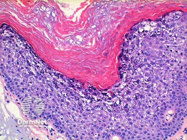
Low power view of histology of epidermolytic hyperkeratosis demonstrates hyperkeratosis and epidermal hyperplasia of varying degrees (Figure 1). The diagnostic features include a characteristic vacuolar degeneration with hypergranulosis of the stratum granulosum and stratum spinosum (Figures 2 and 3).
 pathology
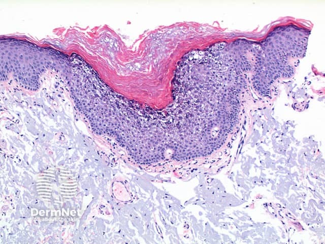
Epidermolytic acanthoma: When the changes of epidermolytic hyperkeratosis are seen forming a solitary lesion. Rarely multiple discrete lesions may be seen in disseminated epidermolytic acanthoma.
Epidermolytic ichthyosis: epidermolytic hyperkeratosis may be seen within biopsies of this generalised congenital condition.
Incidental: Epidermolytic hyperkeratosis may be seen in normal skin adjacent to any skin lesion or dermatosis.
Epidermolytic leukoplakia: is the term used for epidermolytic features arising on a mucosal surface (which is nonkeratinised).
Epidermal naevus variant: epidermolytic hyperkeratosis may be seen within some linear and systematised epidermal naevi.

