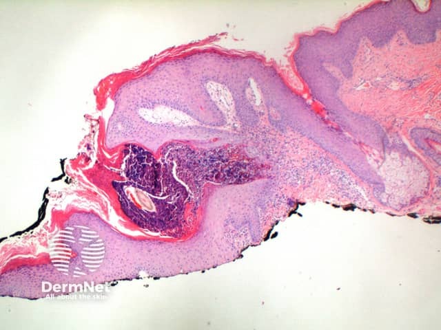
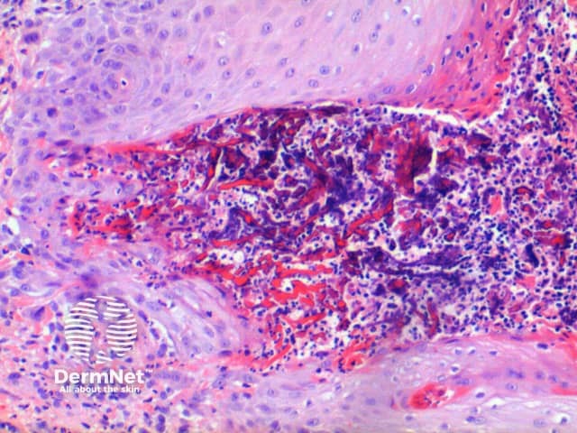
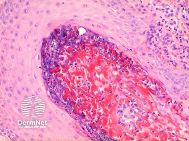
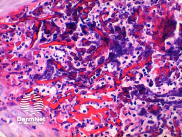
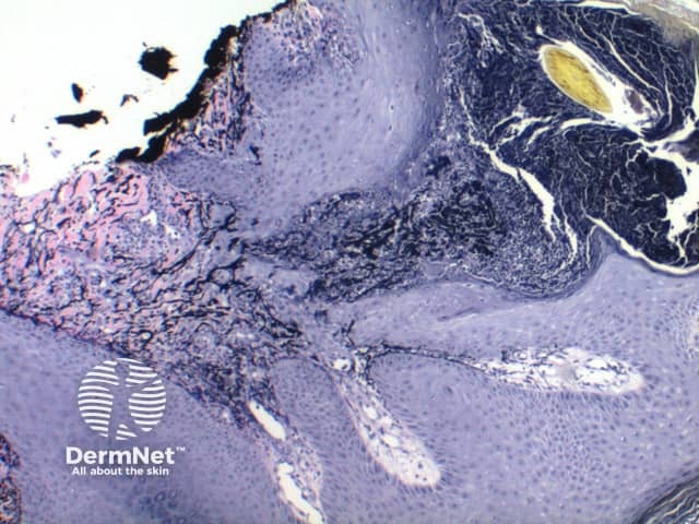
Low power of histology of elastosis perforans serpiginosa demonstrates a column of keratotic debris forming a focal invagination through a hyperplastic epidermis (figure 1). Closer inspection identifies material undergoing transepidermal elimination (figure 2). Brightly eosinophilic fibres are seen within the extruded material, mixed with keratinous debris and a mixed inflammatory cell infiltrate (figures 3, 4).
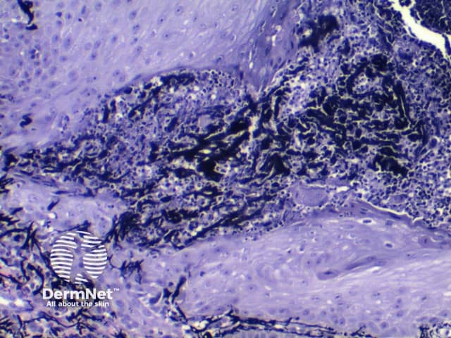
Elastic-Van Gieson (EVG) staining highlights elastic fibres, so in this condition will show black fibres transgressing through the focus of transepidermal elimination (figures 5, 6).
Traumatic collagen perforation: following traumatic erosion of the epidermis, altered collagen fibres in the superficial dermis can appear to be perforating residual epidermis. This is a secondary phenomenon not uncommonly encountered in lesions of prurigo nodularis. Broad areas of epidermal erosion with adjacent epidermal hyperplasia and vertically oriented dermal fibrosis indicating chronic prurigo are clues to the underlying cause. In addition the extruded fibres are collagen and will be negative with EVG staining.
Kyrle disease: in this condition the material being ‘perforated’ is strictly inflammatory debris from the dermis and not collagen or elastic tissue. Overlaps with reactive perforating collagenosis.
Reactive perforating collagenosis: extruded fibres are collagen and will be negative with EVG staining.

