logo

DermNet NZ

Advertisement

Facts about skin from the New Zealand Dermatological Society Incorporated. Topic index: A B C D E F G H I J K L M N O P Q R S T U V W X Y Z

Pigmented follicular cyst – pathology

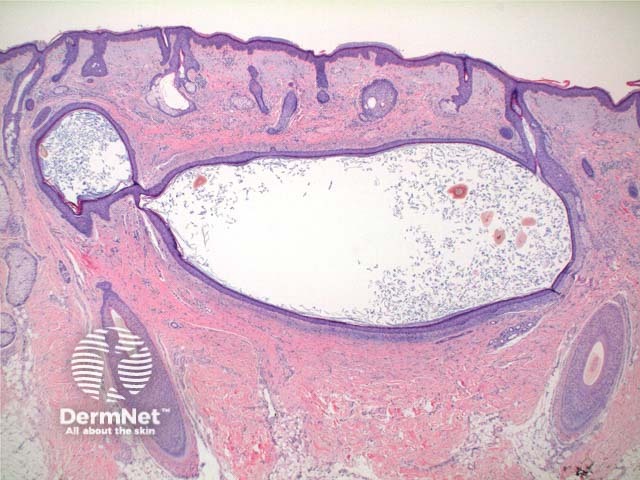
This benign lesion is grouped within the cutaneous cysts

Histology of pigmented follicular cyst

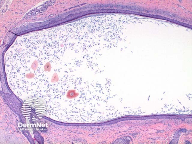
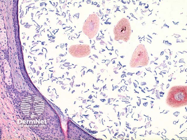
Scanning power view demonstrates a unilocular cystic structure within the dermis (Figure 1). Higher power reveals a thin epithelial lining with a retained granular layer (Figures 2 and 3). Within the cyst cavity are loose keratin fragments and numerous pigmented terminal hair shafts (Figures 2 and 3).

Primary cutaneous amyloidosis – pathology
Figure 1
Primary cutaneous amyloidosis – pathology
Figure 2
Primary cutaneous amyloidosis – pathology
Figure 3
Pigmented follicular cyst – pathology

Differential diagnosis of pigmented follicular cyst

Vellous hair cyst: In this lesion the cyst cavity contains small vellous hairs lacking pigmentation.

Epidermal inclusion cyst: Hair fragments are absent.

Related information

References:

On DermNet NZ:

Other websites:

Books about skin diseases:

See the DermNet NZ bookstore

Author: Dr Ben Tallon, Dermatologist/Dermatopathologist, Tauranga, New Zealand.

DermNet does not provide an on-line consultation service.
If you have any concerns with your skin or its treatment, see a dermatologist for advice.