logo

DermNet NZ

Advertisement

Facts about skin from the New Zealand Dermatological Society Incorporated. Topic index: A B C D E F G H I J K L M N O P Q R S T U V W X Y Z

Lichen nitidus – pathology

Histology of lichen nitidus

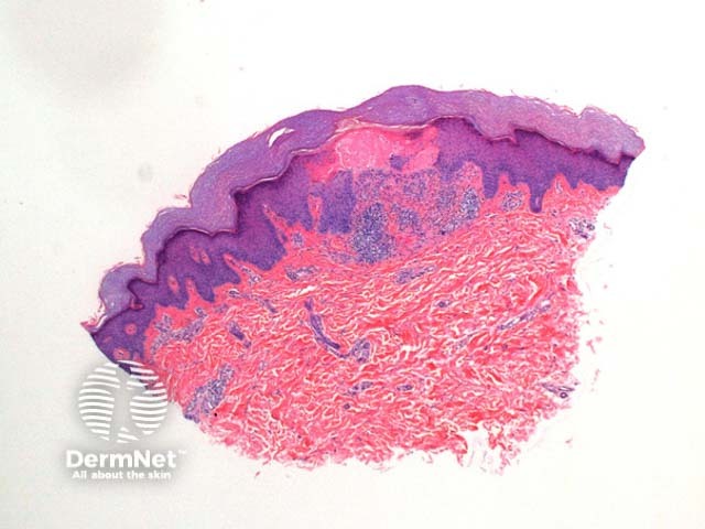
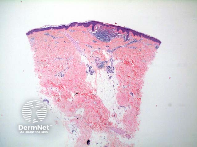
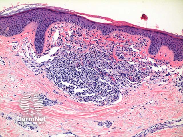
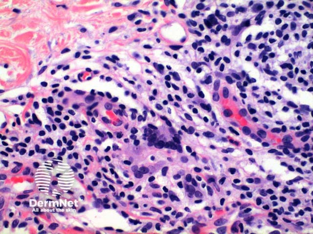
Scanning power view of lichen nitidus identifies a focal dermal inflammatory infiltrate enclosed within collarettes of epidermal acanthosis (Figures 1 and 2). Focal erosion of the epidermis in figure 1 indicates excoriation. Higher power view identifies a well circumscribed lymphohistiocytic infiltrate with multinucleated giant cells (Figures 3 and 4).

Lichen nitidus pathology
Figure 1
Lichen nitidus pathology
Figure 2
Lichen nitidus pathology
Figure 3
Lichen nitidus pathology
Figure 4
Lichen nitidus – pathology

Differential diagnosis of lichen nitidus

Micropapular sarcoidosis: The inflammatory infiltrate is also seen focally within the papillary dermis, but is predominantly histiocytic forming non caseating epithelioid granulomas.

Lichen scrofulosorum: The non caseating granulomas are typically centred on the hair follicles or sweat ducts.

Papular granuloma annulare: Shows localized changes, but otherwise retains the typical infiltrate and collagen changes of granuloma annulare.

Draft 25 September 2011

Related information

References:

On DermNet NZ:

Other websites:

Books about skin diseases:

See the DermNet NZ bookstore

Author: Dr Ben Tallon, Dermatologist/Dermatopathologist, Tauranga, New Zealand.

DermNet does not provide an on-line consultation service.
If you have any concerns with your skin or its treatment, see a dermatologist for advice.