Pleomorphic lipomas are benign tumours, which display atypical cytologic features that may be confused with liposarcoma.
Histology of pleomorphic lipoma
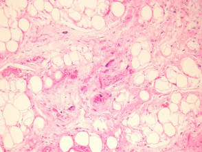
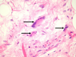
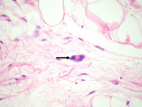
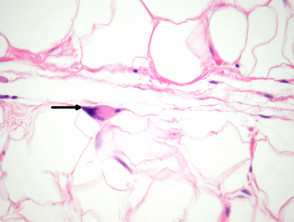
Sections of pleomorphic lipoma show a circumscribed fatty tumour with mature fat admixed with more cellular areas (figure 1). There are often mucinous and spindled areas similar to those seen in spindle cell lipoma. There are intermixed lipoblast-like cells which are giant, may be multinucleated, and often referred to as floret giant cells (figures 2-4, arrows). The nuclei of the giant cells are smudged may be places around the periphery of the cell and have smudged nuclei (otherwise called “floret giant cells”). The cytoplasm is solid and eosinophilic (figures 2-4, arrows).
Pleomorphic lipoma pathology
Special studies for pleomorphic lipoma
The pleomorphic cells are CD34 positive. The fat is S100 positive.
Cytogenetic studies may reveal the lesion shows loss of chromosome 16q or 13q.
Differential diagnosis of pleomorphic lipoma
Spindle cell lipoma – These lesions harbour the same cytogenetic abnormalities as spindle cell lipoma and are regarded by some authorities as the same entity. Pleomophic lipoma displays characteristic floret giant cells.
Liposarcoma – Mitoses favour liposarcoma. Smudgy degenerative changes and floret giant cells are not typically seen in liposarcoma. Cytogenetic studies may be employed in difficult cases. MDM2 and CDK4 gene amplification are features of liposarcoma which can be demonstrated with cytogenetic or immunohistochemical methods.