Talon noir is also called ‘black heel’ or calcaneal petechiae, talon noir is considered to be induced by trauma.
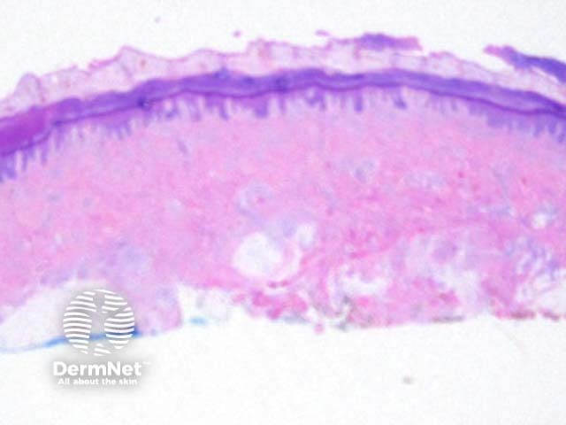
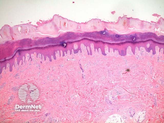
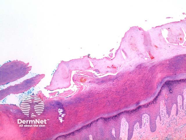
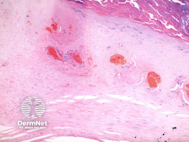
Low power of the histology of talon noir demonstrates acral skin, with notable hyperkeratosis and epidermal acanthosis (Figure 1). The changes are seen within the epidermis. There is inspisated hemorrhage focally and in pools within the stratum corneum (Figure 2, 3 and 4). Telangiectatic vessels and extravasation of erythrocytes may be noted in the papillary dermis (Figure 3).
Verruca vulgaris: Focal hemorrhage is typically seen overlying areas of papillary projection (cap-like hemorrhage), and the additional epidermal changes of a wart make this distinction straight forward in most cases.
The main clinical differential diagnosis is of a melanocytic proliferation (melanoma) or other cutaneous malignancy. Careful inspection is essential so as not to overlook a concurrent melanocytic tumour.

