Introduction Demographics Causes Clinical features Differential diagnoses Diagnosis Treatment Outlook Follow-up
Extramammary Paget disease of the skin is an uncommon intraepithelial adenocarcinoma usually of the anogenital or axillary skin.
Extramammary Paget disease is classified into primary and secondary disease:
Extramammary Paget disease of the skin is differentiated from mammary Paget disease which has a similar appearance involving the nipple or areola, but is a manifestation of an underlying breast cancer.
Extramammary Paget disease generally affects individuals over 50 years of age, with a peak at 65 years. It is more common in Caucasians than other ethnicities. In Asian populations, there is a marked male predominance, in contrast to the female predominance seen in Caucasian populations. In males, Asians and Pacific Islanders are the predominant groups affected, followed by Caucasians, and is rarely seen in African Americans.
The cause of extramammary Paget disease remains poorly understood, however it is most commonly located in apocrine gland-rich skin.
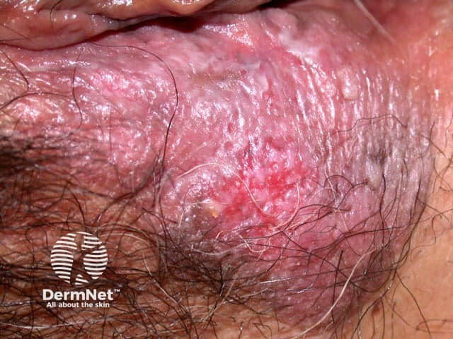
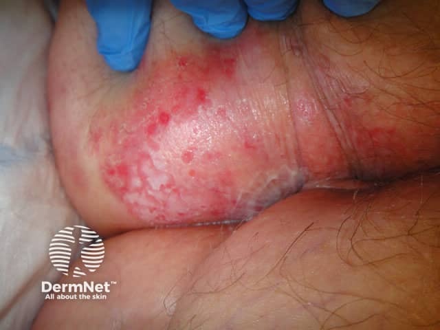
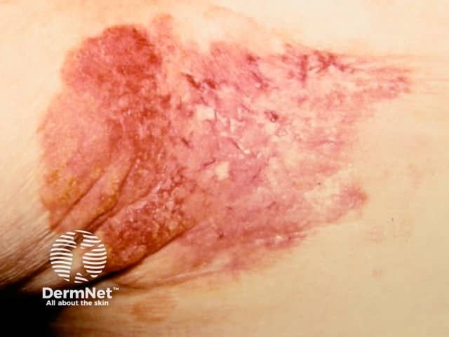
Extramammary Paget disease most commonly presents as an asymmetrical or unilateral, red or pink, scaly plaque on the vulva in women or perianally in men. It is typically slow growing with irregular, poorly defined margins, often leading to misdiagnosis as an inflammatory dermatosis.
Extramammary Paget disease of a non-apocrine-rich region is extremely rare and is termed ectopic extramammary Paget disease. Sites reported have included the scalp, umbilicus, thigh, and face.
Due to the non-specific clinical features, diagnosis of extramammary Paget disease is often delayed by months or years.
Dermoscopy of extramammary Paget disease has been reported to show two unique features — white small round clods with white structureless areas ('cloud-like structureless areas') and thick branching white lines with intermingled white clods ('lava lake structures').
Pigmented extramammary Paget disease can be difficult to distinguish from superficial spreading melanoma, even on dermoscopy. The combination of a linear arrangement of brown globules and a white negative pigment network may be useful clues seen in pigmented extramammary Paget disease.
See more images on Mammary and extramammary Paget disease of the skin images
The differential diagnosis for extramammary Paget disease of the skin includes common inflammatory dermatoses, infections, and other malignancies.
The possibility of extramammary Paget disease must be considered for a chronic skin change in the anogenital region that does not respond to standard topical treatment for dermatitis within 4-6 weeks.
Diagnosis requires a skin biopsy for histopathological confirmation. See extramammary Paget disease pathology.
Once a diagnosis of extramammary Paget disease is made on histology, evaluation for an underlying internal malignancy is required.
Investigations may include:
Surgery, including wide local excision and Mohs micrographic surgery, is the standard treatment for extramammary Paget disease. Surgical procedures tend to be extensive; radical vulvectomy may be required. Despite this, there is a high rate of recurrence due to multifocal disease and poorly defined margins clinically. Mohs micrographic surgery results in a lower recurrence rate compared to wide local excision. Recurrent extramammary Paget disease is usually retreated surgically. Reconstruction may require a skin graft or flap repair.
Sentinel lymph node biopsy may be considered if Paget cells have extended into the reticular dermis or are seen within lymphatics or vascular spaces.
Imiquimod cream is showing good evidence as a useful non-surgical treatment at initial presentation, for recurrent disease, or for those who are not surgical candidates.
Radiotherapy, either alone or as an adjuvant therapy, has also been used with some responses.
Other less successful non-surgical treatments have included:
There are a small number of reports of successful use of trastuzumab for extramammary Paget disease with HER-2 overexpression.
Treatment of metastatic extramammary Paget disease has not been standardised due to the rarity of the condition. Surgery, chemotherapy, radiotherapy, and trastuzumab have been tried.
Any underlying cancer identified on screening will also require appropriate treatment.
In general, patients with extramammary Paget disease have a good prognosis with a 5-year overall survival rate of 75–95%. However, quality of life can be significantly impacted after radical surgery.
Although extramammary Paget disease is usually intraepithelial, it can progress to invasive disease with metastases.
Risk factors associated with a poor prognosis include:
Outcome is also affected by the associated primary underlying cancer.
Due to the high recurrence rate (30–60%), long-term follow-up is recommended to monitor for local disease recurrence, the development of internal malignancy, regional lymphadenopathy, or distant metastasis.

