Introduction Demographics Clinical features Diagnosis Treatment
An oral fibroma is a common benign scar-like reaction to persistent long-standing irritation in the mouth. It is also known as a traumatic fibroma, focal intraoral fibrous hyperplasia, fibrous nodule or oral polyp.
An oral fibroma is most commonly seen in older adults but can occur at any age. It affects 1–2% of adults.
It is usually due to chronic irritation such as:
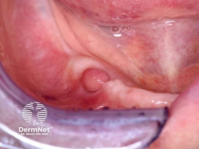
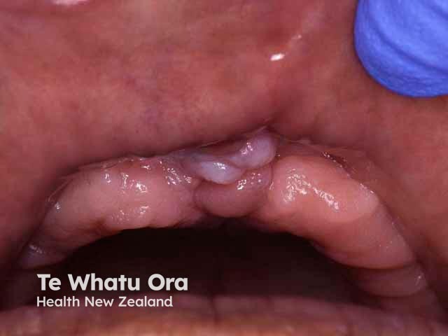
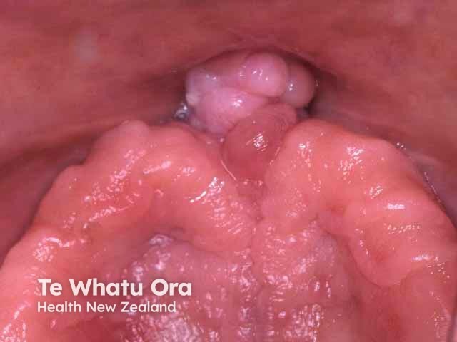
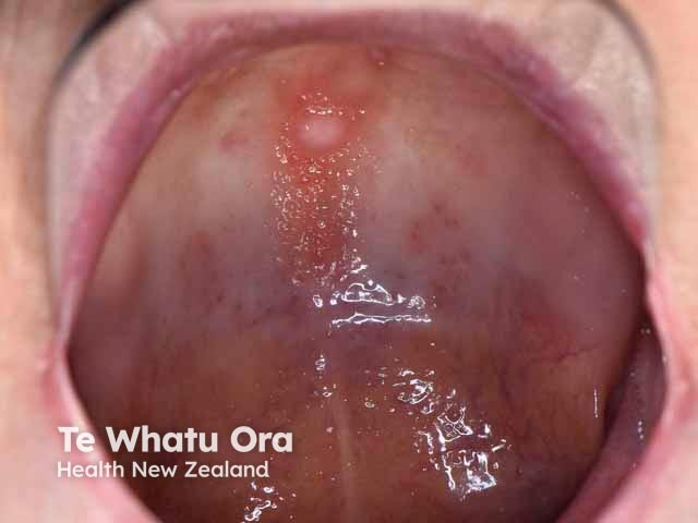
An oral fibroma presents as a firm smooth papule in the mouth. It is usually the same colour as the rest of the mouth lining but is sometimes paler or, if it has bled, may look a dark colour. The surface may be ulcerated due to trauma, or become rough and scaly. It is usually dome-shaped but may be on a short stalk like a polyp (pedunculated). If it has developed under a denture it may be flat with a leaf-like shape.
The commonest location for an oral fibroma is on the inside of the cheek where the upper and lower teeth meet. Other common sites include the sides of the tongue, gums and inside the lower lip.
Apart from the feel and appearance, oral fibromas do not cause any symptoms. Oral fibromas develop over weeks or months to reach a maximum size usually about 1 cm in diameter, but can sometimes be larger.
An oral fibroma is usually a solitary lesion. When there are many lesions, associated diagnoses need to be considered including tuberous sclerosis, Cowden syndrome, familial fibromatosis and fibrotic papillary hyperplasia of the palate.
Oral fibromas do not develop into oral cancer.

In addition to the irritation fibroma, there are a number of other well-recognised types of oral fibroma:
The diagnosis of oral fibroma will be suspected clinically when there are the usual history and examination findings. A biopsy may be taken to exclude other conditions or to remove the lesion. Histology shows typical dense fibrous tissue with relatively few cells. The overlying epithelium may be ulcerated, thinned or thickened.
When treatment is required, the only option is surgical excision of the fibroma with narrow margins. It may recur after surgery if the source of irritation continues. It is therefore also important to manage the source of the irritation. Oral fibromas do not disappear without treatment.

