Introduction Demographics Causes Clinical features Dermoscopic features Complications Diagnosis Differential diagnoses Treatment Outcome
Mammary Paget disease is an uncommon form of breast cancer, comprising 1–3% of all breast cancer presentations. It is also called Paget disease of the breast or nipple.
Mammary Paget disease is nearly always associated with an underlying intraductal breast cancer located near the areola.
Mammary Paget disease is distinguished from extramammary Paget disease seen in the apocrine gland-rich anogenital skin and axillae.
Mammary Paget disease mainly affects women in their 50s and 60s, with a wide age range reported from adolescence to the elderly. Men can also be diagnosed with mammary Paget disease, although this is rare.
There are two main theories proposed for the pathogenesis of mammary Paget disease.
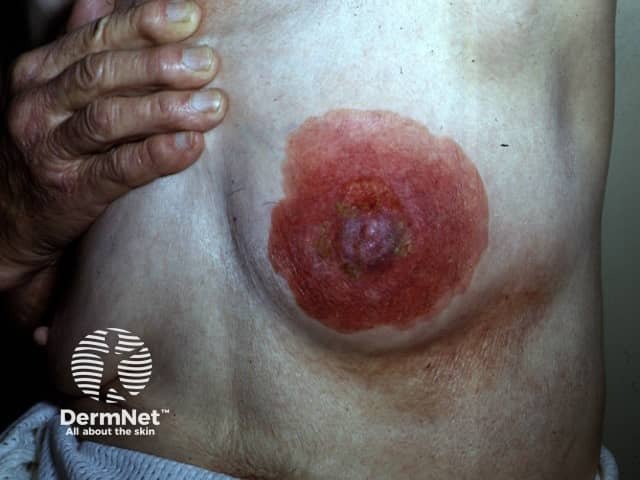
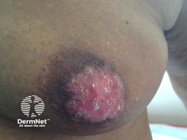
Mammary Paget disease typically starts as an eczema-like change of one nipple, extending with time over the areola, and often associated with an underlying breast lump. It is rarely bilateral. Ectopic and accessory nipples can be affected.
Common cutaneous features of mammary Paget disease include:
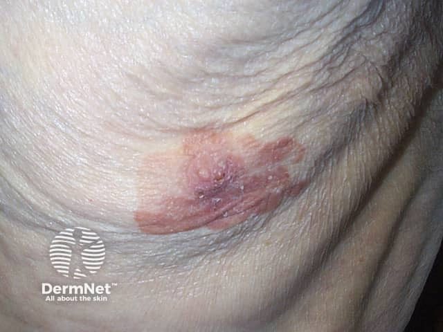
A pigmented variant is well recognised in both males and females, although this presentation is very rare. It can be very difficult to clinically distinguish from melanoma.
See more images in Mammary and extramammary Paget disease of the skin images.
Dermoscopy may be performed to distinguish the lesion from an inflammatory or infective dermatosis or, in the case of pigmented mammary Paget disease, from melanoma.
Dermoscopic features of typical mammary Paget disease have been rarely described, but can include:
Dermoscopy of pigmented mammary Paget disease is reported to be almost indistinguishable from melanoma, with features including:
Mammary Paget disease is almost always a skin sign of an underlying breast cancer. This may be an in-situ or invasive intraductal carcinoma. Metastases to lymph nodes and beyond may be present.
Mammary Paget disease should be suspected clinically when a longstanding, slowly progressive, unilateral nipple change is seen.
A skin biopsy will show the typical foamy Paget cells in the epidermis. In pigmented mammary Paget disease, special stains (eg, CK-7, S-100, Melan A, HMB-45) are required to distinguish Paget cells from malignant melanocytes.
A wedge biopsy of the nipple extends more deeply into the breast tissue and aids diagnosis of the underlying cancer. Immunohistochemistry is used to determine hormone receptor status (oestrogen receptor, progesterone receptor) and human epidermal growth receptor 2 (HER2) that can influence treatment.
Investigations to determine the extent of the associated breast cancer may include ultrasound, mammogram, or MRI.
Mammary Paget disease should be distinguished from:
The usual treatment of mammary Paget disease is surgical excision. Although mastectomy was routine, especially in males, consideration is now given to breast-conserving surgery (removing the nipple, areola, and underlying breast tissue with a margin of healthy tissue). Sentinel lymph node biopsy assesses node involvement. Axillary lymph node dissection may be required if the breast cancer has spread to lymph nodes.
Further treatment may include:
Despite the visible lesion, a delay in presentation or diagnosis is common. On average, diagnosis is made 12 months after onset in women and 8–9 months in men. Prognosis is therefore not as good as for more common forms of breast cancer. It is particularly poor for males, with a 20–30% five-year survival.

