Introduction
Panniculitis is a recognised, but rare complication of α1-antitrypsin (A1AT) deficiency. It is mostly diagnosed in the homozygous PiZZ variant.
Histology of alpha-1-antitrypsin deficiency panniculitis
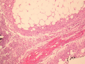
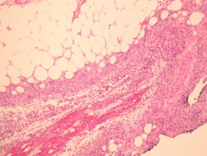
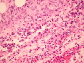
Histopathological examination usually reveals a lobular panniculitis but septal involvement may be seen. In early stages, neutrophils are interstitially arranged between collagen bundles of the deep reticular dermis, followed by a neutrophilic mixed panniculitis with lobular necrosis (figures 1, 2, 3). Vasculitis has been described in association with significant neutrophilic infiltration.
Alpha-1-antitrypsin deficiency panniculitis pathology
Special studies for alpha-1-antitrypsin deficiency panniculitis
Special stains and/or cultures for microorganisms can be helpful if an infectious aetiology is suspected.
Differential diagnosis of alpha-1-antitrypsin deficiency panniculitis
Neutrophilic dermatoses: Clinical correlation can be needed to exclude other neutrophilic dermatoses. Usually patients are known to have alpha-1-antitrypsin deficiency when the panniculitis presents.
Factitial panniculitis: The sections should be polarised to exclude foreign material if a factitial cause is suspected.