What is angiolymphoid hyperplasia with eosinophilia?
Angiolymphoid hyperplasia with eosinophilia is an apparently non-malignant, locally proliferating lesion composed of channels of small blood vessels surrounded by lymphocytes and eosinophils (these are two types of white blood cell). Angiolymphoid hyperplasia with eosinophilia is also known as epithelioid or histiocytoid haemangioma.
Kimura disease is probably distinct from angiolymphoid hyperplasia with eosinophilia. In Kimura disease, the lesions are deeper-seated, with no initial overlying skin lesions. In angiolymphoid hyperplasia with eosinophilia the lesions are smaller and characterised by thick-walled so-called histiocytoid or epithelioid blood vessels.
Angiolymphoid hyperplasia with eosinophilia has been reported from many parts of the world but appears to be particularly common in Japan.
Angiolymphoid hyperplasia with eosinophilia
What are the symptoms of angiolymphoid hyperplasia with eosinophilia?
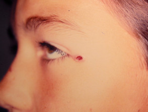
Angiolymphoid hyperplasia with eosinophilia has the following features:
- Affected individuals are commonly young adults, with mean age in mid-30s; it can also affect children
- Clustered small, translucent nodules arise around the ear or the hairline
- Lesions may also involve the inside of the mouth or genitals
- The lesions may be brown or red in colour
- They may be asymptomatic, itchy or painful
- Individual nodules rarely exceed 2–3 cm in diameter, although occasionally they are larger and extend more deeply
- Some cases clear up without treatment, after a variable period of time.
Peripheral blood eosinophilia may be present and is said to be more common in the Kimura variant.
What is the cause of angiolymphoid hyperplasia with eosinophilia?
The cause is unknown, but antigenic stimulation following insect bites has been postulated. It sometimes follows an injury (10%).
Investigations in angiolymphoid hyperplasia with eosinophilia
Skin biopsy shows a poorly organised lesion.
- Clusters of proliferating capillaries are lined with plump, swollen endothelial cells; these are described as epithelioid or histiocytoid because of their appearance.
- A cellular infiltrate composed mainly of lymphocytes and large numbers of eosinophils is seen around the blood vessels.
- Inflammatory cells may penetrate into the lumen of the blood vessels, blocking or rupturing them.
For more details, see angiolymphoid hyperplasia with eosinophils pathology.
A blood test may show increased circulating eosinophil cells (eosinophilia).
Management of angiolymphoid hyperplasia with eosinophilia
If a confident diagnosis of angiolymphoid hyperplasia with eosinophilia is made on a small lesion, it is reasonable to observe the condition for 3-6 months in case spontaneous regression occurs.
Response to active treatment is variable and not always successful.
Simple surgical excision may be done but the lesions tend to recur. Mohs micrographic surgery, including excision of the abnormal vessels at the base of the lesion, may be more effective. There tends to be a lot of bleeding during surgery.
Other possible treatments may include: