What is dermoscopy?
Dermoscopy or dermatoscopy refers to the examination of the skin using skin surface microscopy, and is also called ‘epiluminoscopy’ and ‘epiluminescent microscopy’. Derm(at)oscopy is mainly used to evaluate pigmented skin lesions. In experienced hands it can make it easier to diagnose melanoma.
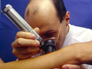
Dermoscopy requires a high quality magnifying lens and a powerful lighting system (a dermatoscope). See below for more information on dermatoscopes.
Computer software can be used to archive dermoscopy images and allow expert diagnosis and reporting (mole mapping). Smart programs may aid in diagnosis by comparing the new image with stored cases with typical features of benign and malignant pigmented skin lesions.
Dermoscopy
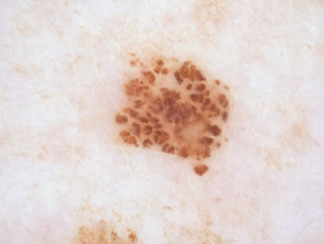
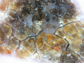
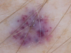
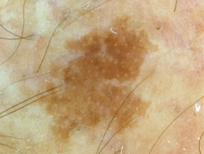
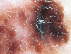
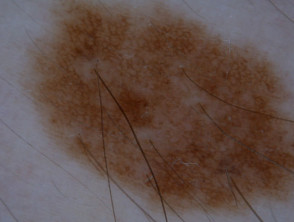
Dermatoscopic features of pigmented lesions
Using dermoscopy, the pigmentation of the lesion is evaluated in terms of colour(s) and structure.
Colours found in pigmented skin lesions include black, brown, red, blue, grey, yellow and white.
The colours of dermoscopy
Characteristics of the dermatoscopic structure of the skin lesions include:
- Symmetry or asymmetry
- Homogeny/uniformity (sameness) or heterogeny (structural differences across the lesion)
- Distribution of pigment: brown lines, dots, clods and structureless areas
- Skin surface keratin: small white cysts, crypts, fissures
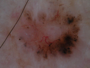
- Vascular morphology and pattern: regular or irregular
- Border of the lesion: fading, sharply cut off or radial streaks
- Presence of ulceration
There are specific dermoscopic patterns that aid in the diagnosis of the following pigmented skin lesions:
- Melanoma
- Moles (benign melanocytic naevus)
- Freckles (lentigos)
- Atypical naevi
- Blue naevi
- Seborrhoeic keratosis
- Pigmented basal cell carcinoma
- Haemangioma
Skin lesions viewed by dermoscopy
Dermoscopy may also be used for detailed skin surface examination in some other circumstances, for example:
- Non-pigmented skin lesions
- Finding a scabies mite within a burrow
- Locating a splinter
- Examining nail fold capillaries in cutaneous lupus erythematosus or systemic sclerosis
- Distinguishing certain skin conditions such as lichen planus from others such as psoriasis or eczema
- Evaluating hair loss (using trichoscopy)
Dermatoscopes
Used widely by dermatologists, plastic surgeons, and general practitioners, dermatoscopes allow the user to perform skin surface microscopy. This enables the practitioner to examine skin structures and patterns.
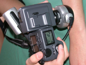
There are several different dermatoscopes and these are either conventional standalone devices, or smartphone attachable. When choosing a dermatoscope, there are some important things to consider:
- Compatibility - smartphones and cameras
- Polarisation - polarised, linear-polarised, unpolarised, or variable
- Contact or Non-contact viewing - or both
- Magnification - eg, 10x, 16x, or 40x
- Battery - disposable, rechargeable, time to charge, time of continuous operation
- Cost
See also our Dermatoscope comparative image gallery.