What is erythema nodosum?
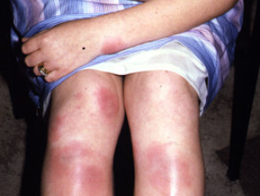
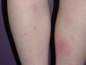
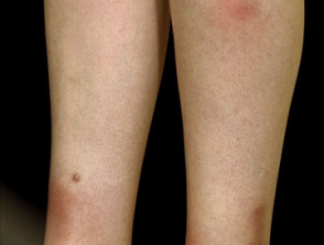
Erythema nodosum is a type of panniculitis, an inflammatory disorder affecting subcutaneous fat. It presents as tender red nodules on the anterior shins. Less commonly, they affect the thighs and forearms [1–3].
Erythema nodosum
See more images of erythema nodosum.
Who gets erythema nodosum?
Erythema nodosum can occur in all ethnicities, sexes, and ages, but is most common in women between the ages of 25 and 40 [4]. It is 3–6 times more common in women than in men except before puberty when the incidence is the same in both sexes [5].
What are the causes of erythema nodosum?
Erythema nodosum is a hypersensitivity reaction of unknown cause in up to 55% of patients [6]. In other cases, it is associated with an identified infection, drug, inflammatory condition, or malignancy [7].
- Throat infections (streptococcal disease or viral infection)
- Primary tuberculosis (TB), a rare cause in New Zealand
- Yersinia infection; this causes diarrhoea and abdominal pain
- Chlamydia infection
- Fungal infection: histoplasmosis, coccidioidomycosis
- Parasitic infection: amoebiasis, giardiasis
Other viral and bacterial diseases associated with erythema nodosum include herpes simplex, viral hepatitis, human immunodeficiency virus (HIV) infection, Campylobacter infection, and Salmonella infection.
Drugs (3–10%)
- Sulfonamide
- Amoxicillin
- Oral contraceptive
- Non-steroidal anti-inflammatory drugs
- Bromide
- Salicylate
- Iodide
- Gold salt
Inflammatory
- Inflammatory bowel disease (ulcerative colitis or Crohn disease)
- Sarcoidosis (11–-25%); X-ray shows bilateral hilar adenopathy in Löfgren syndrome
- Malignancy
- Lymphoma
- Leukaemia
- Behçet disease
Others
- Pregnancy (2-5%)
What are the clinical features of erythema nodosum?
Erythema nodosum presents with tender bilateral erythematous subcutaneous nodules 3–20 cm in diameter erupting over one to several weeks. They are accompanied by fever and joint pain. In 50% the ankle is swollen and painful for up to several weeks. The knees and other joints can also be affected [8].
Common clinical findings [9-12]
- The nodules are found on the anterior lower legs, knees and arms and rarely on the face and neck.
- They are ill-defined, warm, oval, round or arciform, and without ulceration
- The nodules are initially bright to deep red.
- They spontaneously resolve within eight weeks, through a violaceous, brownish, or yellowish/green bruise-like appearance known as erythema contusiformis.
Erythema nodosum does not cause permanent scarring.
What are the complications of erythema nodosum?
Erythema nodosum has few known complications and lesions usually resolve spontaneously. A rare complication is encapsulated fat necrosis, or ‘mobile encapsulated lipoma’ [14].
How is erythema nodosum diagnosed?
Erythema nodosum is primarily a clinical diagnosis confirmed by laboratory tests and histopathology [8]. The pathology of erythema nodosum shows inflammation primarily of the septa between the subcutaneous fat lobules without vasculitis [15].
Supporting investigations [4,7]
Appropriate tests may include:
- Complete blood count with differential, C-reactive protein levels (infectious and inflammatory causes)
- Chest X-ray (tuberculosis and sarcoidosis)
- Throat swab and anti-streptolysin O and streptodornase serology (streptococcal infection)
- Viral serology (preferably two samples at four-week intervals)
- Stool culture and evaluation for ova and parasites in patients with gastrointestinal symptoms
- Mantoux test or QuantiFERON gold (tests for TB).
- Deep incisional or excisional skin biopsy.
What is the differential diagnosis for erythema nodosum?
A range of causes of panniculitis should be considered in a patient with subcutaneous nodules, especially if lesions are not located on the legs, there is ulceration, or symptoms last longer than eight weeks.
Panniculitis can be predominantly septal (inflammation between lobules) or lobular (inflammatory cells within subcutaneous fat lobules) [16]. Mixed septal and lobular inflammation can occur.
Nodules due to predominantly septal panniculitis include:
- Various forms of scleroderma
- Medium vessel vasculitis, for example, due to polyarteritis nodosa in which there are tender subcutaneous nodules associated with ulceration, necrosis, livedo racemosa, fever, joint pain, myalgia, and peripheral neuropathy
- Necrobiosis lipoidica
- Eosinophilic panniculitis
- Rheumatoid nodule.
Nodules due to predominantly lobular panniculitis include:
- Connective tissue disease (eg, panniculitis associated with cutaneous lupus erythematosus)
- Erythema nodosum leprosum (type 2 lepra reaction due to leprosy)
- Pancreatic panniculitis in which subcutaneous nodules may become ulcerated or fluctuant. Laboratory tests reveal elevated lipase, amylase and trypsin levels
- Traumatic panniculitis
- Nodular vasculitis/erythema induratum in which ulcerated, draining nodules affect the posterior calves
- Lipodermatosclerosis, which is a consequence of venous insufficiency
- Infection of the subcutaneous fat with bacteria, mycobacteria or fungi, which may cause ulcerated, fluctuant, and draining abscesses
- Malignant infiltration.
What is the treatment for erythema nodosum?
Erythema nodosum is treated based on the underlying disease. An underlying infection should be treated.
- Pain management may include extended rest, colchicine (1–2 mg/day), NSAIDs (non-steroidal anti-inflammatory drugs), and venous compression therapy [4].
- Systemic corticosteroids (1 mg/kg daily until resolution of erythema nodosum) may be prescribed if infection, sepsis, and malignancy have been ruled out [9,11].
- Oral potassium iodide as a supersaturated solution (400–900 mg/day) may be prescribed for one month if available [17].
What is the outcome for erythema nodosum?
Erythema nodosum follows a relatively benign and favourable course. It is important to recognise the underlying cause, if any, and initiate symptomatic treatment [4]. Most cases resolve within days to weeks. Relapses may occur in approximately one-third of cases erythema nodosum may become a chronic or persistent disorder lasting for 6 months and occasionally for years [13].


