Introduction Properties Types of lasers Treatment of skin conditions Other uses Laser safety Adverse effects
Note to our readers. This page is overdue for an update; there have been significant changes in laser technology since it was originally written.
The first lasers used to treat skin conditions occurred over 40 years ago. Argon and carbon dioxide (CO2) lasers were commonly used to treat benign vascular birthmarks such as port-wine stains and haemangiomas. Although these birthmarks could be effectively lightened, a side effect was the unacceptably high rate of scar formation. In the last 20 years, advances in laser technology have revolutionised their use in the treatment of many skin conditions and congenital defects, including vascular and pigmented lesions, and the removal of tattoos, scars and wrinkles. There is a spectrum of laser and light technologies available for skin resurfacing and rejuvenation.
‘Laser’ is an acronym: light amplification by the stimulated emission of radiation.
Lasers are sources of high-intensity light with the following properties:
Laser light can be accurately focused into small spots with very high energy.
The light is produced within an optical cavity containing a medium, which may be a gas (eg, argon, krypton, carbon dioxide), liquid (eg, dye) or solid (eg, ruby, neodymium:yttrium-aluminium-garnet, alexandrite). The process involves excitation of the molecules of the laser medium, which results in the release of a photon of light as it returns to a stable state. Each medium produces a specific wavelength of light, which may be within the visible spectrum (violet 400 through to red 700nm) or infrared spectrum (more than 700 nm).
Vascular skin lesions contain oxygenated haemoglobin, which strongly absorbs visible light at 418, 542 and 577 nm, whereas pigmented skin lesions contain melanin, which has a broad range of absorption in the visible and infrared wavebands. Infrared lasers are broadly destructive because they are absorbed by water in and between skin cells (these are composed of 70-90% water).
The aim is to destroy the target cells and not to harm the surrounding tissue. Short pulses reduce the amount that the damaged cells heat, thereby reducing thermal injury that could result in scarring. Automated scanners aim to reduce the chance of overlapping treatment areas.
There are several types of lasers used in skin laser surgery. Older laser technologies such as the continuous wave (CW) lasers of CO2 and argon have been replaced with quasi-CW mode lasers and pulsed laser systems. Picosecond lasers have very short pulses.
The wavelength peaks of the laser light, pulse durations and how the target skin tissue absorbs this, determine the clinical applications of the laser types.
CW lasers emit a constant beam of light with long exposure durations
Quasi-CW lasers shutter the CW beam into short segments, producing interrupted emissions of constant laser energy
Laser source |
Wavelength peaks |
|||
|---|---|---|---|---|
532 nm |
||||
Copper bromide/vapour |
510/578 nm |
|||
Argon-pumped tunable dye (APTD) |
577/585 nm |
|||
Krypton |
568 nm |
|||
Pulsed* lasers emit high-energy laser light in ultrashort pulse durations with relatively long intervening periods between each pulse
Laser source |
Wavelength peaks |
|||
|---|---|---|---|---|
Pulsed dye laser (PDL) |
585–595 nm |
|||
QS ruby |
694 nm |
|||
QS alexandrite |
755 nm |
|||
1064 nm |
||||
2940 nm |
||||
10,600 nm |
||||
*Pulsed laser systems may be either long-pulsed such as PDL with pulse durations ranging from 450ms to 40millisec, or very short-pulsed (5-100ns) such as the quality-switched (QS) lasers.
Picosecond lasers uses very short pulse durations to target endogenous pigmentation
Laser source |
Wavelength peaks |
|||
|---|---|---|---|---|
532/1064 nm |
||||
755 nm |
||||
Lasers have been used successfully to treat a variety of vascular lesions including superficial vascular malformations (port-wine stains), facial telangiectases, haemangiomas, pyogenic granulomas, Kaposi sarcoma and poikiloderma of Civatte. Lasers that have been used to treat these conditions include argon, APTD, KTP, krypton, copper vapour, copper bromide, pulsed dye lasers and Nd:YAG. Argon (CW) causes a high degree of non-specific thermal injury and scarring and is now largely replaced by yellow-light quasi-CW and pulsed laser therapies.
The pulsed dye laser is considered the laser of choice for most vascular lesions because of its superior clinical efficacy and low-risk profile. It has a large spot size (5 to 10mm) allowing large lesions to be treated quickly. Side effects include postoperative bruising (purpura) that may last 1-2 weeks and transient pigmentary changes. Crusting, textural changes and scarring are rarely seen.
The new V-beam features provide ultra-long pulse duration so energy directed at the target blood vessels over a longer period, resulting in more uniform blood vessel damage reducing the purpura seen with the earlier pulse dye lasers. The addition of dynamic cooling increases comfort during treatment enabling higher fluencies (energy) to be delivered safely and effectively, so fewer treatments are required.
Vascular malformations associated with smaller more superficial blood vessels respond better to treatment than deeper larger vessels (more often arising in older individuals). It is, therefore, best to begin treatment early. Fading by 80% occurs after 8 to 10 treatments on average. Further treatment may be necessary if the lesion recurs.
Treatment with quasi-CW lasers also produce effective outcomes, but they may be associated with higher incidences of scarring and textural changes. The most common side effects include mild erythema, oedema, and transient crusting.
Non-laser intense pulsed light devices can also be used for treating vascular lesions.

Melanin-specific, high energy, QS laser systems can successfully lighten or eradicate a variety of pigmented lesions. Pigmented lesions that are treatable include freckles and birthmarks including some congenital melanocytic naevi, blue naevi, naevi of Ota/Ito and Becker naevi. The short pulse laser systems effectively treat the lesions by confining their energy to the melanosomes, which are the tiny granules containing melanin inside the pigment cells. The results of laser treatment depend on the depth of the melanin and the colour of the lesion and are to some degree unpredictable. Superficially located pigment is best treated with shorter wavelength lasers while removal of deeper pigment requires longer wavelength lasers that penetrate to greater tissue depths. Caution is needed with laser therapy in skin of colour, as permanent hypopigmentation and depigmentation may occur. Successfully treated lesions may recur.
Before any laser treatment of pigmented lesions, any lesion with atypical features should be biopsied to rule out malignancy. The treatment of congenital melanocytic naevi is a controversial issue. The long-term effect of using lasers on promoting melanoma is not known, but the treatment is thought to be low risk.
The QS laser systems can selectively destroy the tattoo pigment without causing much damage to the surrounding skin. The modified pigment is removed from the skin by scavenging white blood cells, tissue macrophages. The choice of laser depends on the colour, depth and chemical nature of the tattoo ink. Two to ten treatments are often necessary. Yellow, orange and green are the most difficult colours to remove.
As with other laser treatments, pigmentary and textural changes including scars may occur.
Picosecond Nd:YAG and alexandrite lasers have been found to remove exogenous pigment more effectively than QS lasers.
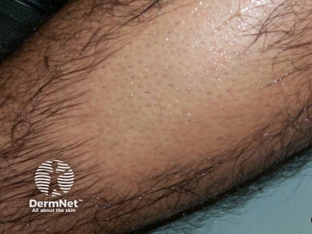
Lasers can be used to remove excessive and cosmetically disabling hair due to hypertrichosis or hirsutism. Laser treatments remove dark hair quickly, and it may take 3 to 6 months before regrowth is evident. Several treatment cycles are required with the spacing between treatments depends on the body area being treated. Laser treatments are less painful and much quicker than electrolysis. Complications are rare but superficial burns, pigmentary changes and even scarring may occur. Increased growth of fine dark hair in untreated areas close to the treated ones has been reported. Both increased and reduced localised sweating have been reported after treatment.
Suitable devices include long-pulsed ruby and alexandrite lasers, a diode (810nm), millisecond Nd:YAG and non-laser intense pulsed light.

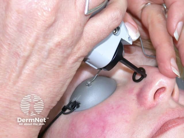
Facial laser resurfacing uses high-energy, pulsed and scanned lasers.
Pulsed CO2 and erbium:YAG lasers have been successful in reducing and removing facial wrinkles, acne scars and sun-damaged skin. High-energy, pulsed, and scanned CO2 laser is generally considered the gold standard against which all other facial rejuvenation systems are compared. Typically a 50% improvement is found in patients receiving CO2 laser treatment. Side effects of treatment include post-operative tenderness, redness, swelling and scarring. The redness and tenderness last several weeks, while new skin grows over the area where the damaged skin has been removed by the laser treatments (ablative laser systems). Secondary skin infection including reactivation of herpes is also a potential problem until healing occurs. Extreme caution is needed when treating darker-skinned individuals as a permanent loss, or variable pigmentation may occur longterm.
Erbium:YAG produces similar results and side effects to the CO2 laser. Despite their side effect profile and long recovery time these ablative laser systems, when used properly, can produce excellent results.
Recently non-ablative lasers have been used for dermal modelling; 'non-ablative' refers to heating the dermal collagen while avoiding damage to the surface skin cells (epidermis) by cooling it. Multiple treatments are required to smooth the skin.
Keloids and hypertrophic scars are difficult to eradicate, and traditional treatments are not always successful. Vaporising lasers (CO2 and erbium:YAG) have been useful as an alternative to conventional surgery. More recently PDL has been used to improve hypertrophic scars and keloids. This may require multiple treatment sessions or the simultaneous use of intralesional injections to gain good results. The PDL has been reported to reduce the redness as well as improving the texture and flexibility of the scar.
Lasers are sometimes used to remove viral warts by vaporisation (CO2 laser) or destruction of the dermal blood vessels (PDL), but the evidence would suggest that this is no more effective than standard wart paints or even waiting for spontaneous clearance.
The CO2 laser can be used to remove a variety of skin lesions including seborrhoeic keratoses and skin cancers by vaporisation or in cutting mode. However, conventional surgery or electrosurgery can also be used and is generally less expensive.
Violet-blue metal halide light (407-420 nm) has been used to treat acne because it has a toxic effect on the acne bacteria, Cutibacterium acnes.
The Excimer laser uses noble gas and halogen to produce ultraviolet radiation (308 nm) that will clear psoriasis plaques. However, the small spot size and the tendency to cause blistering makes treatment time-consuming and difficult to perform.
Safety precautions will depend on which laser system is used and in what setting. They should include:
Laser treatments are burns. The following adverse effects may occur:
Medscape Reference

