Histology Special stains Histological variants Differential diagnoses
Leiomyosarcoma is a malignant tumour of smooth muscle. It rarely affects the skin.
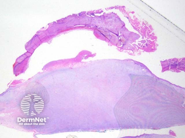
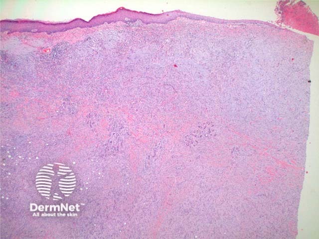
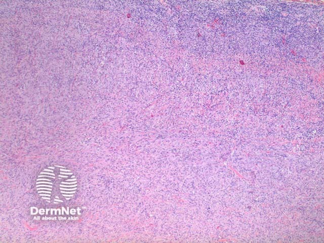
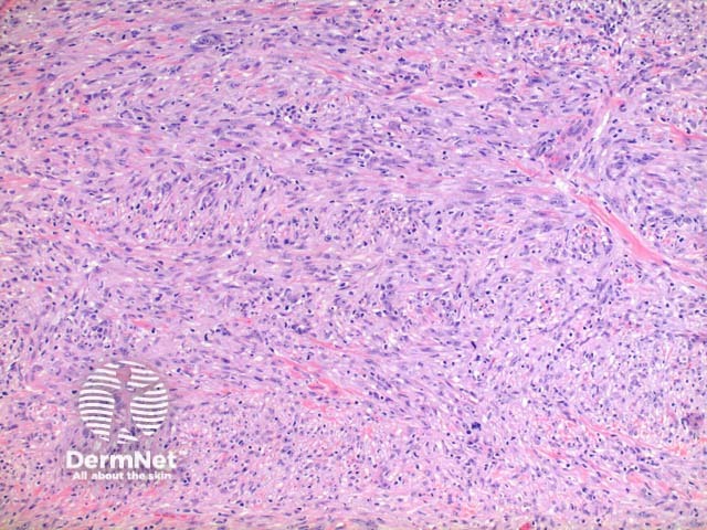
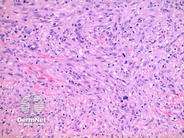
Scanning power view of histology of leiomyosarcoma shows a poorly circumscribed tumour nodule which may be dermal based in the less common dermal leiomyosarcoma (Figure 1) or deeply infiltrating in the subcutaneous form. The tumour is comprised of a spindle cell proliferation forming rough bundles and fascicles (Figures 2 and 3). High power demonstrates spindle cells with cigar shaped nuclei with prominent cytologic atypia and mitotic figures (Figures 4 and 5).
Immunoperoxidase staining is positive for muscle markers SMA(smooth muscle actin), HHF35 (pan muscle actin), h-caldesmon and Desmin. Negative Desmin staining is seen infrequently. A rare number of cases have demonstrated cytokeratin or EMA (epithelial membrane antigen) staining.
Atypical leiomyoma: This lesion likely falls on a spectrum between leiomyoma and leiomyosarcoma. Reliable criteria to exclude malignancy in cutaneous leiomyosarcomas are lacking but uterine discriminating features reported include: size less than 5.5cm, mitotic count less than 7 per 10 high power fields in the presence of significant nuclear atypia and lack or tumour cell necrosis.
A mitotically active leiomyoma is used to describe a lesion lacking significant nuclear atypia or tumour necrosis, but with between 5 and 15 mitoses per high power field.
Other malignant spindle cell tumours to consider include spindle cell squamous cell carcinoma, malignant peripheral nerve sheath tumour, spindle cell melanoma, angiosarcoma or atypical fibroxanthoma. Immunoperoxidase staining is typically required to differentiate between these lesions.

