What is melanoma of the nail unit?
Melanoma of the nail unit is usually a variant of acral lentiginous melanoma, a malignant melanoma arising from the palms of the hands and soles of the feet. Other rare forms of melanoma that may arise in the nail unit are nodular melanoma or desmoplastic melanoma.
Melanoma of the nail unit most often affects the great toe and thumbnail, accounting for 75–90% of cases. However, any nail on the finger or toe may be involved.
The term includes:
- Subungual melanoma (melanoma originating from the nail matrix)
- Ungual melanoma (melanoma originating from under the nail plate)
- Periungual melanoma (melanoma originating from the skin beside the nail plate).
Melanoma of the nail unit
Who gets melanoma of the nail unit?
Melanoma of the nail unit is rare. While occurring equally in all racial groups, it accounts for around 0.7–3.5% of malignant melanomas in white-skinned populations and up to 75% of dark-skinned and Asian populations.
It is the most common type of melanoma diagnosed in deeply pigmented individuals, probably due to this population's low incidence of cutaneous melanoma, due to the melanin pigment protection from ultraviolet (UV) radiation.
It is most often diagnosed in the 60 to 70-year-old age group.
Melanoma management is an evolving area. For up-to-date recommendations, refer to the Australian Cancer Council Clinical practice guidelines for the diagnosis and management of melanoma.
What causes melanoma of the nail unit?
In contrast to cutaneous melanoma, melanoma of the nail does not appear to be related to sun exposure. Melanoma of the nail unit originates from activation and proliferation of melanin producing melanocytes of the nail matrix.
Injury or trauma may be a factor, accounting for the greater incidence in the big toe and thumb (75–90% of cases).
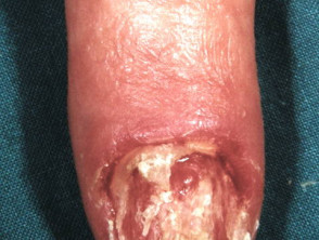
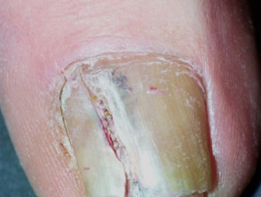
What are the clinical features of melanoma of the nail unit?
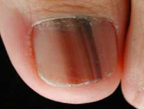
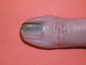
Melanoma of the nail unit most often starts as a narrow brown to black pigmented band, visible on the length of a single nail plate (melanonychia). It is most prevalent in the nail of the thumb or big toe. It is not easy to differentiate from a benign lentigo or naevus (mole) in its early stages. However, over weeks to months, the pigment band:
- Becomes wider (>3 mm), especially at its proximal end (cuticle)
- Becomes more irregular in pigmentation (light and dark brown)
- Borders become irregular or blurred
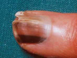
- Extends to involve the skin of the proximal or lateral nail fold (periungual pigmentation), also known as the “Hutchinson sign”, which is historically considered a sign of nail melanoma
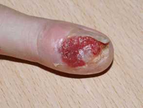
- May form a nodule, ulcerate, or bleed
- May present as a mass under the nail plate, lifting it (onycholysis), or look like a wart (verrucous)
- May cause nail plate dystrophy (thinning, cracking or distortion of the nail plate)
- May become painful if there is bone involvement.
Important considerations
- Melanoma of the nail unit is hypomelanotic or amelanotic in up to 50% of all cases.
- The Hutchinson sign, although valuable, is not always an accurate predictor of melanoma.
- Periungual pigmentation is present in many benign disorders, such as Bowen disease of the nail unit.
- Nail pigmentation may be an illusion, especially in children with nail matrix nevi, due to transparency of the nail folds showing through the pigment.
- "Pseudo-Hutchinson’s sign" is a phrase that encompasses these simulants of Hutchinson's sign.
Melanoma of the nail unit
How do clinical features vary in differing types of skin?
Melanoma of the nail unit arises in people of all skin colour and race.
It is important to note that while longitudinal melanonychia only occurs in around 1% of white-skinned individuals, it is very common in dark-skinned races, occurring in an estimated 100% of African-Americans by the age of 50 and around 20% of Asians. This may lead to misdiagnosis or delayed diagnosis of nail unit melanoma, and therefore poorer outcomes.
What are the complications of melanoma of the nail unit?
- Metastatic spread of the disease to other body parts.
- The vertical growth phase can cause nail dystrophy and ulceration.
- Cosmetic deformity.
How is melanoma of the nail unit diagnosed?
Melanoma of the nail unit diagnosis requires:
History
A thorough history to establish the onset, progression, and possible triggers. Clues to the aetiology should be sought, including medical and drug history, digital trauma, and any external exposures.
Examination
A thorough examination of all nails looking for:
- Number of nails involved
- Pigmented band (width, colour and homogeneity)
- Signs of trauma
- Nail dystrophy or subungual nail lesions.
Dermoscopic examination (onychoscopy)
Dermoscopy of the nail plate may help with an early clinical diagnosis of melanoma of the nail and help rule out lesions not requiring further examination. However, clinicians should maintain a low threshold of suspicion for performing a biopsy for histopathologic examination. Dermoscopic clues for nail unit melanoma include:
- The width of the pigmented band occupying more than two-thirds of the nail plate
- Grey and black colours
- Irregularly pigmented lines (in their colour, spacing, thickness, and parallelism)
- Hutchinson sign and micro-Hutchinson sign (skin pigmentation noted on dermoscopy but not on clinical examination)
- Nail dystrophy
- Granular pigmentation (found in 40% of melanomas and only in 3.5% of benign lesions).
The ABCDEF mnemonic is helpful for the assessment of pigmented nail lesions:
- A: Age (50–70 years old); African, Japanese, Chinese, and Native American heritage
- B: Brown-black pigmented band ≥3mm with blurred borders
- C: Change or lack of change despite treatment in the nail band or nail morphology
- D: Digit most commonly involved (thumb, big toe, or index finger)
- E: Extension of pigment into the skin surrounding the nail (Hutchinson sign)
- F: Family or personal history of melanoma or dysplastic nevus (atypical mole)
Nail biopsy
Definitive diagnosis requires a biopsy of the nail matrix and nail bed. Any clinical features suspicious of melanoma of the nail unit warrants a biopsy.
Some experts suggest avoiding nail matrix biopsies in children where possible (unless the band is getting larger or darker).
Nail histopathology
Histopathologic examination is the gold standard for diagnosing nail melanoma. Differentiating early melanoma of the nail from benign lesions is often challenging due to similar features. The pathologist will report if the melanoma is invasive and the tissue level invaded. Common subtypes are acral lentiginous, followed by nodular and desmoplastic.
What is the differential diagnosis for melanoma of the nail unit?
- Subungual haematoma/haemorrhage
- Exogenous discolouration (colour caused by external factors)
- Splinter haemorrhage
- Fungal melanonychia
- Longitudinal erythronychia
- Onychopapilloma
- Pigmented onychomatricoma
- Subungal glomus tumour
- Naevi (moles)
- Lentigo
- Subungual squamous cell carcinoma (SCC)
- Systemic illness (including systemic lupus erythematosus, scleroderma)
- Drug-induced pigmentation
- Ethnic-type pigmentation (can often involve multiple digits)
What is the treatment for melanoma of the nail unit?
General measures
The management plan for melanoma of the nail unit will usually require a multidisciplinary melanoma team to direct further investigations and treatment. The mainstay of management of melanoma of any kind is excision.
Specific measures
- Melanoma in situ: may be managed more conservatively by wide surgical excision of the entire nail apparatus with margin control.
- Invasive melanoma: must be removed surgically. Traditionally this has required digit amputation. Digit sparing approaches to preserve the bone and joints, such as wide local excision and Mohs micrographic surgery, may be considered for early melanoma of the nail unit. However, this requires careful patient selection and expertise about the anatomy and pathology of the nail unit.
- Metastatic melanoma: may benefit from management with newer immunotherapy agents.
- A sentinel node biopsy may be performed to determine spread to local lymph nodes.
Melanoma management is an evolving area. Please refer to your local guidelines or speak with a medical specialist for up-to-date treatment protocols.
What is the outcome for melanoma of the nail unit?
Melanoma of the nail unit is an uncommon form of melanoma with a worse prognosis than other cutaneous melanomas. The main factor associated with the risk of metastatic melanoma and death is the thickness of the melanoma at the time of primary tumour excision. It is often diagnosed late, particularly when the toe is involved, and may have spread at the time of diagnosis.
According to the American Cancer Society, the 5-year survival rate for cutaneous melanoma varies between 15–97% depending on stage. Melanoma of the nail unit may have a worse prognosis compared with cutaneous melanoma, but this may be related to the late presentation. A meta-analysis of mortality rates for melanoma of the nail unit reported a five-year survival of 77%.