Myxofibrosarcoma is an uncommon soft tissue tumour, typically presenting in the lower limb girdle of the elderly. It is a malignant tumour with high risk of local recurrence.
Histology of myxofibrosarcoma
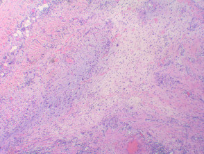
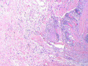
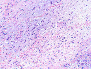
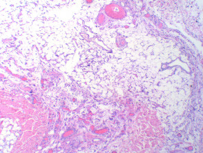
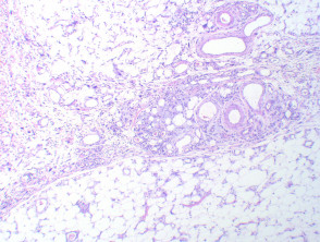
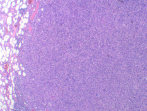
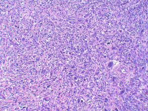
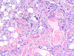
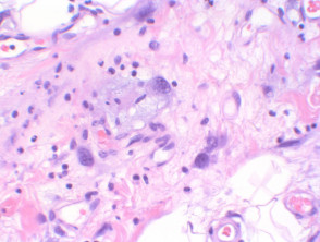
Myxofibrosarcoma presents as a multinodular tumour, with a variably cellular spindle cell proliferation in a myxoid background (figures 1, 2). Cellular atypia with nuclear pleomorphism and enlarged hyperchromatic nuclei is seen at least focally (figure 3). The characteristic feature is the presence of delicate curvilinear vessels (figures 4,5).
Grading falls on a spectrum based on interpretation of cellularity.
- Low grade: Hypocellular with no solid areas.
- Intermediate grade: Myxoid with increased cellularity.
- High grade: Solid tumour with prominent pleomorphism (figures 6,7)
Special stains
In general immunohistochemistry is unhelpful in myxofibrosarcoma, but is positive for vimentin, and sometimes weakly positive for smooth muscle actin and CD34.
Myxofibrosarcoma variants
Epithelioid: Cellular areas morphologically epithelioid. Behave as high grade, and are prone to increased local recurrence and risk of metastasis.
Differential diagnosis
Low grade fibromyxoid sarcoma: Typically paucicellular with alternating fibrous and myxoid areas. Nuclear pleomorphism is mild or absent.
Myxoid liposarcoma: This tumour has lipoblasts and lacks significant pleomorphism. Vacuoles within the lipoblasts will be clear, as opposed to myxofibrosarcoma, where the vacuoles typically contain fine myxoid material (pseudolipoblasts) (figures 8,9). Genetics will show a t(12;16) or t(12;22) translocation, while MDM2 is frequently negative.
Myxoid dermatofibrosarcoma protuberans: Storiform pattern with CD34 positive immunostaining.
Malignant fibrous histiocytoma: This entity behaves like, and likely is synonymous with, high grade myxofibrosarcoma.