DermNet provides Google Translate, a free machine translation service. Note that this may not provide an exact translation in all languages

Translate

Onycholysis

Authors: Vanessa Ngan, Staff Writer, 2003. Updated: Dr Fleur Weiqian Kong, Honorary Dermatology Registrar, Mater Adult Hospital, Brisbane QLD, Australia; Dr James Muir, Dermatologist, Mater Adult Hospital, Brisbane QLD, Australia. Copy edited by Gus Mitchell. December 2020.


toc-icon

What is onycholysis?

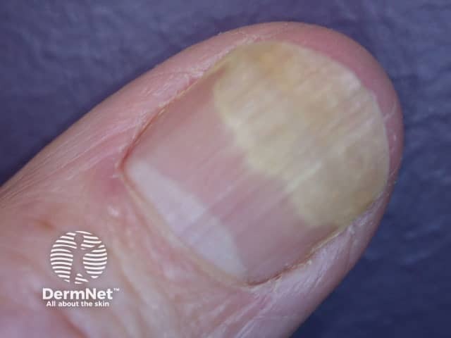
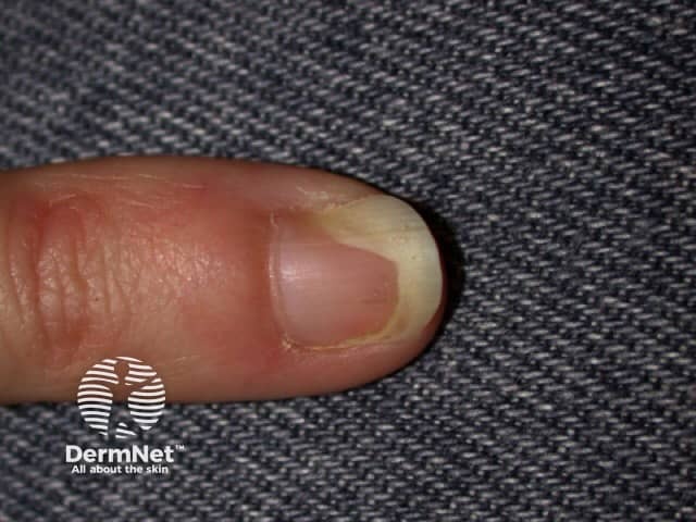
Onycholysis is a common nail disorder in which the nail plate has separated from the nailbed typically resulting in a well-defined area of white opaque nail. It may be idiopathic or secondary to trauma, skin disease, nail infections, tumours, or systemic events. Photo-onycholysis is due to ingestion of a photosensitiser, such as a medication [see Drug-induced photosensitivity].

Onycholysis

Who gets onycholysis?

Onycholysis can affect both sexes, all ages and races. It is most frequently seen in adult women.

What causes onycholysis?

Onycholysis can be primary (idiopathic, unknown cause) or secondary to one of many causes. Some examples are listed in Table 1.

Cause Examples
Traumatic

Repetitive trauma, including manicure

Prolonged immersion in water

Chemical exposure including nail cosmetics

Foreign body implantation

Skin disease Psoriasis

Hand dermatitis

Lichen planus
Infection Dermatophytes and yeasts eg, T. rubrum, C. albicans

Scabies
Systemic disease Endocrine eg, hypo- and hyperthyroidism, diabetes mellitus, pregnancy

Infiltrative eg, amyloidosis, sarcoidosis

Malignancy eg, multiple myeloma, Sezary syndrome

Photosensitivity eg, porphyrias

Yellow nail syndrome
Medication Drug-induced photosensitivity eg, antibiotics including tetracyclines, NSAIDs, psoralens, and oral retinoids

Chemotherapeutic agents eg, taxanes
Others Hereditary distal onycholysis (MIM 164800)

What are the clinical features of onycholysis?

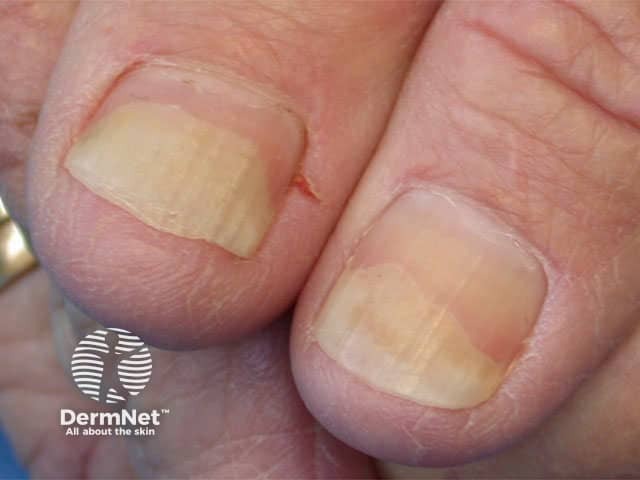
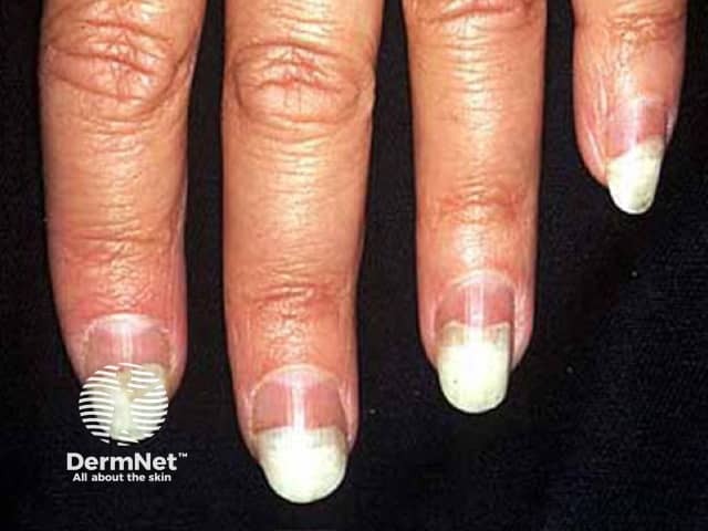
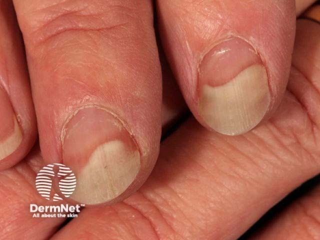
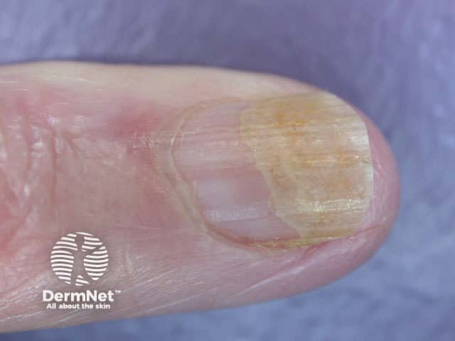
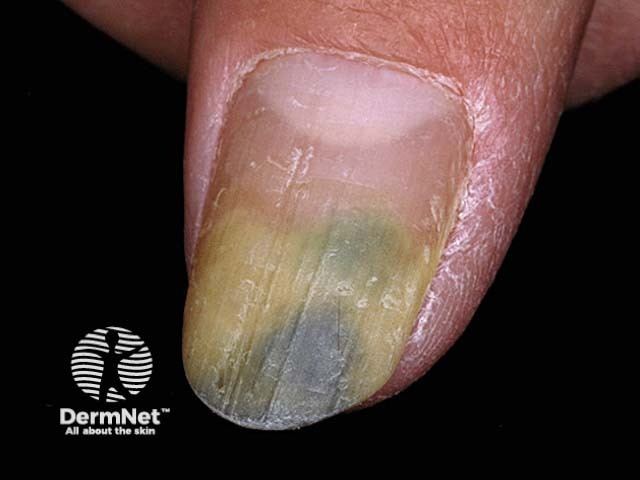
Onycholysis can affect a single nail or multiple fingernails and/or toenails. The distal part of the nail is most commonly affected lifting the free edge; sometimes the nail may detach laterally or proximally. Oil spot sign is an island of onycholysis under a nail.

Clinical features can include the following signs.

  • An irregular but sharply defined border between the pink portion of the nail and the white edge of the lifted detached nail.
  • The detached nail is usually white and opaque compared to the transparent normal nail that appears pink.
  • Lateral/proximal onycholysis and oil spot sign are a yellow-pink colour rather than white.
  • The nail surface may be normal or pitted, indented, or crumbly.
  • Thickened hyperkeratotic skin may accumulate under the detached nail.
  • Subungual haemorrhage can occur in acute-onset onycholysis.
  • Onycholysis is usually painless, but may be painful if acute or inflammatory.

Onycholysis in dermatoses

What are the complications of onycholysis?

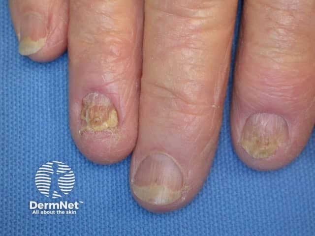
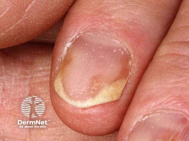
Onycholysis predisposes to secondary infection under the nail, most commonly with Candida albicans and Pseudomonas aeruginosa, resulting in discolouration of the nail.

Onycholysis can be cosmetically unacceptable, especially for people who work with their hands in public view.

Complications of onycholysis

How is onycholysis diagnosed?

Onycholysis is a clinical diagnosis with the cause often obvious on history and examination. Investigations may be required if the cause is not apparent.

What is the differential diagnosis for onycholysis?

Onycholysis may persist or progress due to:

Onycholysis should be distinguished from leukonychia (white nail), including Terry nail, in which the nail remains attached but appears white and opaque.

What is the treatment for onycholysis?

The detached portion of the nail will not reattach. The aim of treatment is for the new nail growth to remain attached to the underlying nailbed.

General measures

  • Clip the affected portion of the nail and keep the nail(s) short with frequent trimming.
  • Minimise activities that traumatise the nail and nailbed.
  • Avoid potential irritants such as nail enamel, enamel remover, solvents, and detergents.
  • Wear gloves, including light cotton gloves under vinyl gloves for wet work.
  • Tape nail to the underlying digit.
  • Use of antimicrobial soaks, such as dilute vinegar, to minimise the risk of secondary infection.

Specific measures

Specific treatment of onycholysis depends on the cause. This may mean ceasing or changing a medication, specific treatment of a nail infection, or appropriate treatment for an associated systemic condition or dermatosis.

What is the outcome for onycholysis?

Onycholysis of short duration and of known origin can recover with appropriate treatment. Fingernails take 4–6 months to fully regrow; toenails take twice as long. The longer onycholysis persists the less likely new nail growth will reattach due to permanent damage (cornification) of the underlying nailbed (‘disappearing nailbed’).


Bibliography

  • Bazex J, Baran R, Monbrun F, Grigorieff-Larrue N, Marguery MC. Hereditary distal onycholysis--a case report. Clin Exp Dermatol 1990;15(2):146–8. doi:10.1111/j.1365-2230.1990.tb02054.x. PubMed
  • Byrne JP, Boss JM, Dawber RP. Contraceptive pill-induced porphyria cutanea tarda presenting with onycholysis of the finger nails. Postgrad Med J. 1976;52(610):535–8. doi:10.1136/pgmj.52.610.535. PubMed Central
  • Chiriac A, Brzezinski P, Foia L, Marincu I. Chloronychia: green nail syndrome caused by Pseudomonas aeruginosa in elderly persons. Clin Interv Aging. 2015;10:265–7. doi:10.2147/CIA.S75525. PubMed Central
  • Damasco FM, Geskin LJ, Akilov OE. Nail changes in Sézary syndrome: a single-center study and review of the literature. J Cutan Med Surg. 2019;23(4):380–7. doi:10.1177/1203475419839937. PubMed
  • Daniel CR 3rd, Iorizzo M, Piraccini BM, Tosti A. Simple onycholysis. Cutis. 2011;87(5):226–8. PubMed
  • De Berker DAR, Richert B, Baran R. Acquired disorders of the nails and nail unit. In: Griffiths C, Barker J, Bleiker T, Chalmers R, Creamer D (eds). Rook’s Textbook of Dermatology [4 volumes], 9th edn. Wiley Blackwell, 2016: 95.9–10.
  • Erpolat S, Eser A, Kaygusuz I, Balci H, Kosus A, Kosus N. Nail alterations during pregnancy: a clinical study. Int J Dermatol. 2016;55(10):1172–5. doi:10.1111/ijd.13316. PubMed
  • Hay RJ, Baran R, Moore MK, Wilkinson JD. Candida onychomycosis--an evaluation of the role of Candida species in nail disease. Br J Dermatol. 1988;118(1):47–58. doi:10.1111/j.1365-2133.1988.tb01749.x.
  • Huang KL, Lin KY, Huang TW, et al. Prophylactic management for taxane-induced nail toxicity: a systematic review and meta-analysis. Eur J Cancer Care (Engl). 2019;28(5):e13118. doi:10.1111/ecc.13118. PubMed
  • Tempark T, Lekwuttikarn R, Chatproedprai S, Wananukul S. Nail scabies: an unusual presentation often overlooked and mistreated. J Trop Pediatr 2017;63(2):155–9. doi:10.1093/tropej/fmw058. PubMed
  • Zaias N, Escovar SX, Zaiac MN. Finger and toenail onycholysis. J Eur Acad Dermatol Venereol. 2015;29(5):848–53. doi:10.1111/jdv.12862. PubMed

On DermNet

Other websites

Books about skin diseases


Related information

Sign up to the newsletter