Angiolymphoid hyperplasia with eosinophilia is an apparently non-malignant, locally proliferating lesion composed of channels of small blood vessels surrounded by lymphocytes and eosinophils (these are two types of white blood cell). Angiolymphoid hyperplasia with eosinophilia is also known as epithelioid or histiocytoid haemangioma.
Kimura disease is probably distinct from angiolymphoid hyperplasia with eosinophilia. In Kimura disease, the lesions are deeper-seated, with no initial overlying skin lesions. In angiolymphoid hyperplasia with eosinophilia the lesions are smaller and characterised by thick-walled so-called histiocytoid or epithelioid blood vessels.
Angiolymphoid hyperplasia with eosinophilia has been reported from many parts of the world but appears to be particularly common in Japan.
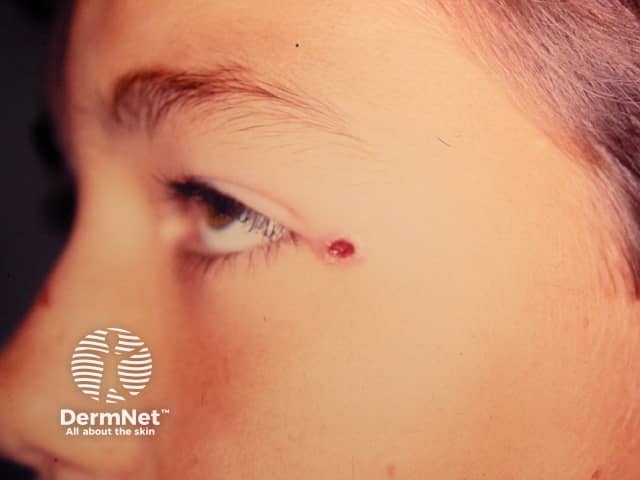
Angiolymphoid hyperplasia with eosinophilia has the following features:
Peripheral blood eosinophilia may be present and is said to be more common in the Kimura variant.
The cause is unknown, but antigenic stimulation following insect bites has been postulated. It sometimes follows an injury (10%).
Skin biopsy shows a poorly organised lesion.
For more details, see angiolymphoid hyperplasia with eosinophils pathology.
A blood test may show increased circulating eosinophil cells (eosinophilia).
If a confident diagnosis of angiolymphoid hyperplasia with eosinophilia is made on a small lesion, it is reasonable to observe the condition for 3-6 months in case spontaneous regression occurs.
Response to active treatment is variable and not always successful.
Simple surgical excision may be done but the lesions tend to recur. Mohs micrographic surgery, including excision of the abnormal vessels at the base of the lesion, may be more effective. There tends to be a lot of bleeding during surgery.
Other possible treatments may include:

