Why do we need laboratory testing for viral infections?
Various tests are carried out in a laboratory to establish or confirm the diagnosis of a viral skin infection. Although a thorough history and examination of the patient are vital, laboratory tests can help the clinician to make a diagnosis.
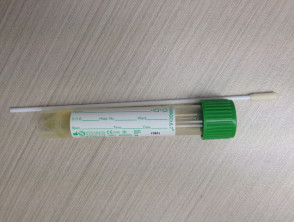
What is a viral skin swab?
A viral skin swab is a sterile implement lightly rubbed against a visible skin lesion or vesicle. The swab is then sent to the laboratory in a viral transport medium for further viral cell culture and virus identification.
What organisms can viral swabs detect?
A viral swab from an external skin lesion or mucosal surface can detect:
- Herpes simplex virus (HSV) types 1 and 2 (cause of herpes simplex infections including genital herpes)
- Varicella-zoster virus (VZV) (cause of chickenpox and shingles)
In addition, a viral swab from oral skin mucosa can detect:
- Epstein-Barr virus (EBV) (the cause of infectious mononuclosis)
- Coxsackievirus A16 (one of the causes of hand, foot and mouth disease)
Viral skin swab
What is a viral cell culture?
Viral cell culture places samples into suitable cell cultures that the virus being tested can infect. When the cells show particular changes the culture is positive. Viral cell culture can identify HSV, VZV, morbillivirus (cause of measles) and other viruses.
What blood tests are needed?
Blood tests for the investigation of viral infections include:
- Full blood count — a viral infection may raise or reduce the white cell count; atypical lymphocytes may be reported
- C-reactive protein (CRP) — this is elevated but usually less than 50 in a viral infection (CRP is a marker of inflammation anywhere in the body and is not a specific test for viral infections)
- Procalcitonin — this is negative. It is a blood test marker for generalised sepsis due to bacterial infection
- Serology — two samples of blood tested 10 days apart to determine immune response to a particular organism.
Polymerase chain reaction (PCR)
PCR involves isolating and amplifying lengths of DNA and then detecting known genetic sequences of microorganisms. Skin swabs can be used to identify:
- Herpes simplex virus (HSV)
- Varicella-zoster virus (VZV)
- Coxsackieviruses
- Human papillomavirus (HPV) (the cause of viral warts and some kinds of cancer)
- Orf virus (a parapox virus from sheep/goats that causes orf).
Enzyme-linked immunosorbent assay (ELISA)
ELISA can test for specific organisms either by detecting the antigen during a current infection or more commonly, antiviral antibody. The detection of the antibody confirms contact with the virus at some time but it is not necessarily the reason for a current infection.
Most commonly detected viral antibodies are to:
- Varicella-zoster virus (VSV)
- Human immunodeficiency virus (HIV)
- Hepatitis B and C virus (in serum), the main causes of viral hepatitis.
Skin biopsy
A skin biopsy may be useful in the diagnosis of viral infection. A viral cytopathic effect may be observed on histopathology or Tzanck smear, or specific features characteristic of the particular infection may be present.
What is electron microscopy?
Electron microscopy involves directing a beam of electrons at a sample producing an image. It can provide much higher resolution images than a standard light microscope. It is rarely used in practice. However it can be useful to identify atypical and rare viral infections in the immunosuppressed patients presenting with unusual skin lesions.
What is resistance testing in viruses?
Resistance testing in viruses is done to aid the clinician as to what antiviral medications may or may not be effective for infection. As viruses have the potential to mutate rapidly, much faster than bacteria or fungi, this presents a therapeutic challenge for the clinician, as treatment with antiviral medications may start to fail. The most noteworthy is HIV antiretroviral (ARV) resistance, which confers a great deal of risk to the patient if the medications stop working.
Sequence analysis is performed most commonly at the genomic level, which looks directly at the HIV genome to see to which antiretroviral drugs that strain of HIV is already resistant and therefore which drugs will not work. Less commonly, this analysis is performed at the phenotypic level, where the patient's HIV virus is subjected to antiretroviral medication in the laboratory to see how well it responds.