DermNet provides Google Translate, a free machine translation service. Note that this may not provide an exact translation in all languages

Translate

Quiz
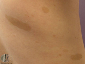
Brown skin lesions – 12 cases

Brown skin colour is most often due to melanin, a protein produced in the epidermis by melanocytes. Various dermopathies have characteristic patterns of pigmentation.

For each of the twelve cases, study the image(s) and then answer the questions. You can click on the image to view a larger version if required.

Each case should take approximately 2 minutes to complete. There is a list of suggested further reading material at the end of the quiz.

Case 1

Café au lait macules

Melanocytic naevi may be congenital or appear in early childhood (congenital type); there are several distinct varieties. The café-au-lait macule is a well-demarcated light brown patch. Histologically, these have nests of naevus cells along the dermoepidermal junction. The naevus cells have variable-sized melanosomes including macromelanosomes. Neurofibromatosis is characterised by multiple café au lait macules, but these may also be seen sporadically and as a feature of other neurocutaneous disorders, including Albright’s syndrome and tuberous sclerosis. A café-au-lait macule with darker spots is known as speckled lentiginous naevus or naevus spilus – this is usually solitary.

Mark question

Sign up to the newsletter