What is an atypical mycobacterial infection?
Atypical mycobacterial infections are infections caused by a species of mycobacterium other than Mycobacterium tuberculosis, the causative bacteria of pulmonary TB and extrapulmonary TB including cutaneous TB; and Mycobacterium leprae, the cause of leprosy.
Atypical mycobacteria may cause many different types of infections, which are divided into the following four clinical syndromes:
- Pulmonary disease
- Lymphadenitis
- Skin and soft tissue disease
- Disseminated disease
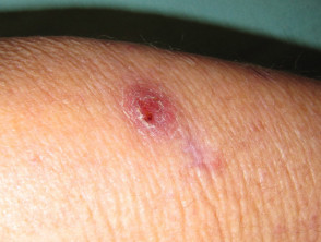
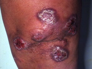
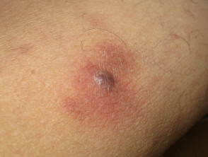
Skin infection tends to result in crusted nodules and plaques. Abscesses may develop in skin and bone infection.
Cutaneous atypical mycobacterial infection
See more mycobacterial infection images
What causes an atypical mycobacterial infection?
There are many different species of mycobacterium. To date at least 30 species of mycobacteria that do not cause tuberculosis or leprosy have been identified. Some of those causing atypical mycobacterial infections include:
- Mycobacterium avium-intracellulare
- Mycobacterium kansasii
- Mycobacterium marinum
- Mycobacterium ulcerans
- Mycobacterium chelonae
- Mycobacterium fortuitum
- Mycobacterium abscessus
Mycobacterium avium-intracellulare and Mycobacterium kansasii primarily cause lung disease similar to pulmonary TB, whilst Mycobacterium marinum, Mycobacterium ulcerans, Mycobacterium fortuitum and Mycobacterium chelonae cause skin infections.
What are the clinical features of an atypical mycobacterial infection?
The clinical features of atypical mycobacterial infection depend on the infecting mycobacteria.
Mycobacterium avium-intracellulare
- Also known as MAC (Mycobacterium avium complex)
- Most common non-tuberculous mycobacterial infection associated with AIDS
- Symptoms include fever, swollen lymph nodes, diarrhoea, fatigue, weight loss and shortness of breath
- May develop into pulmonary MAC
- Skin lesions are uncommon and non-specific
Mycobacterium kansasii
- May cause a chronic infection of the lungs similar to pulmonary TB
- Second most common non-tuberculous mycobacterial infection associated with AIDS
- Symptoms include fever, swollen lymph nodes and lung crackles and wheezing
- Skin lesions may occur either alone or as part of a more widespread disease
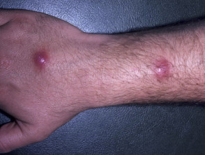
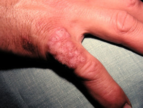
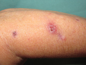
Mycobacterium marinum
- Also known as fish tank granuloma, swimming pool granuloma
- Uncommon infection that occurs most often in people with recreational or occupational exposure to contaminated freshwater or saltwater
- Usually, a single lump or pustule that breaks down to form a crusty sore or abscess
- Other lumps may occur around the initial lesion, particularly along the lines of lymphatic drainage (sporotrichoid forms)
- Most often affects elbows, knees, top of feet, knuckles or fingers
- Multiple lesions and widespread disease may occur in immunocompromised patients
- Rarely causes red, swollen and tender joints (bursitis, tenosynovitis, arthritis, osteomyelitis)
Mycobacterium marinum
See more mycobacterial infection images
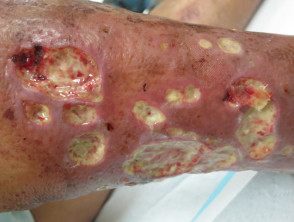
Mycobacterium ulcerans
- Also known as Buruli ulcer, Kumasi, Bairnsdale ulcer
- Infection most common in Central and West Africa around areas of lush vegetation and swamps but may also occur in Australia
- Found in fish, amphibians and the water
- Solitary, painless and sometimes itchy nodule of 1–2 cm develops about 7–14 days after infection through broken skin
- Over one to two months the nodule may break down to form a shallow ulcer that spreads rapidly and may involve up to 15% of the patient's skin surface
- Ulceration and necrosis are due to a toxin, mycolactone
- Severe infections may destroy blood vessels, nerves, and invade bone
- Cultures are very slow to grow
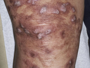
Mycobacterium chelonae
- Worldwide distribution: found in tap water and other water sources
- Can follow tattoos, trauma or surgery
- Mainly affects middle-aged individuals with some degree of immune suppression
- May cause fever, lung disease, joint infection, eye disease and other organ infections
- May result in a non-healing wound, subcutaneous nodule, cellulitis or abscess
- Immunosuppression may cause disseminated lesions throughout the body
Mycobacterium chelonae
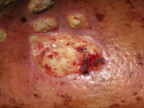
Mycobacterium abscessus
- Subspecies of M. chelonae
- Found in water, soil, dust, animals
- Rarely causes illness in humans but can be difficult to diagnose and treat
- Can cause skin infection after puncture wounds, tattoos, skin trauma or surgery
- May cause lung infection and disseminated infection in immunosuppressed people
Mycobacterium abscessus
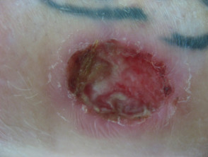
Mycobacterium fortuitum
- Worldwide distribution: found in natural and processed water sources, sewage and dirt
- Local cutaneous disease, osteomyelitis, joint infections and ocular disease may occur after trauma (including tattoos, shaving followed by footbath)
- Often affects healthy young patients
- Severe immunosuppression, especially AIDS, may cause disseminated skin and soft tissue lesions
- Often the cause of wound and surgical-site infections from contaminated water sources
- Causes a non-healing ulcerative skin lesion, furunculosis and/or subcutaneous nodules
See more mycobacterial infection images
How is an atypical mycobacterial infection diagnosed?
Atypical mycobacteria are diagnosed on the culture of tissue. Specific conditions are required, such as cool temperature, so the laboratory must be informed of the clinician's suspicion of this diagnosis. The infections have specific pathological features on skin biopsy.
Other diagnostic tools used include radiographic imaging studies and more recently, polymerase chain reaction (PCR) testing on swabs of ulcers or tissue biopsies.
What is the treatment of atypical mycobacterial infection?
Treatment of atypical mycobacterial infections depends upon the infecting organism and the severity of the infection. In most cases a course of antibiotics is necessary. These include rifampicin, ethambutol, isoniazid, minocycline, ciprofloxacin, clarithromycin, azithromycin and cotrimoxazole. Usually, treatment consists of a combination of drugs.
Consider the following points when treating atypical mycobacterial infections with antibiotics:
- Mycobacterium marinum species are often resistant to isoniazid, streptomycin, pyrazinamide, and para-aminosalicylic acid. Effective antimicrobials include tetracyclines, fluoroquinolones, macrolides (eg, clarithromycin), rifampicin and sulfonamides (cotrimoxazole). Treatment should be for at least 4–6 weeks, and sometimes up to two months.
- Mycobacterium kansasii should be treated with at least 3 drugs for 12–18 months. One of the drugs must be rifampicin, which is still the cornerstone of treatment for these infections.
- Mycobacterium chelonae and M fortuitum are best treated with clarithromycin or azithromycin in localised infections, particularly if used with surgical debridement. Disseminated infections require combination treatment, usually a macrolide and an aminoglycoside eg, combinations of amikacin, tobramycin, imipenem, clarithromycin.
- Treatment of Mycobacterium ulcerans is most successful if treatment is started in lesions less than 6 months old with a diameter less than 10 cm. Rifampicin and streptomycin are the currently recommended antibiotics.
- Surgery is used as an adjunct to antibiotic treatment in patients with severe infection. Most lesions eventually spontaneously heal after 6–9 months but may leave behind extensive scarring and disfigurement.
- AIDS patients on HIV protease inhibitor drugs cannot be treated with rifampicin because rifampicin significantly increases the breakdown of these drugs. Rifabutin is a suitable alternative.
Surgical removal of infected lymph nodes and aggressive debridement of infected skin lesions is sometimes necessary. In severe cases, skin grafts may be necessary to repair the surgical wound.
Some infections will heal spontaneously, leaving a scar (which is often very unsightly).