What is a basal cell carcinoma?
A basal cell carcinoma (BCC) is a keratinocytic or non-melanoma skin cancer formed by uncontrolled growth and replication of basal cells in the lower levels of the epidermis. A BCC is sometimes referred to as a rodent ulcer or basalioma.
What is skin of colour?
Skin of colour is a subjective term referring to natural skin pigmentation ‘darker’ than white (ie, brown or black skin). When compared against a graded assessment of skin colour types such as the Fitzpatrick scale, ‘skin of colour’ may refer to skin classified as type IV or higher [1]. In some contexts, ‘skin of colour’ is also used to describe the skin type of a non-white ethnic group including those of African, Asian, South American, Pacific Island, Maori, Middle Eastern, and Hispanic descent [1]. See ethnic dermatology.
Basal cell carcinoma in skin of colour
Who gets basal cell carcinoma?
Basal cell carcinomas (BCC) are among the most commonly diagnosed cancers worldwide and accounts for 65–75% of skin cancers [2]. It is the most common form of skin cancer seen in Caucasians, Asians, and Hispanics, and the second most common form of skin cancer seen in African Americans and Asian Indians [3].
Incidence data
Reported incidence rates of BCC vary across different studies and periods, although the BCC incidence rates per 100,000 population across the last 50 years include:
- 0.32–6.4 in Chinese
- 0.7–1.4 in Asian Indians
- 1–2 in African Americans
- 6 in New Zealand Maori (for non-melanoma skin cancers in general) [2–4].
The incidence rates of BCC globally are increasing, and this trend is significantly more prominent in white-skinned people, particularly in older age groups and in women [5,6]. Most diagnoses of BCC occur at over 50 years of age and peak at around 70–80 years of age in both men and women, regardless of skin colour [5].
Risk factors for basal cell carcinoma
Basal cell carcinoma is more common in individuals with:
- Previous BCC or other skin cancers (squamous cell carcinoma, melanoma)
- Repeated prior episodes of sunburn
- Outdoor occupation or outdoor recreation, particularly during childhood
- Previous cutaneous injury, thermal burn, or dermatological disease (eg, cutaneous lupus, sebaceous naevus)
- Inherited syndromes: including basal cell naevus syndrome (Gorlin syndrome), Bazex–Dupré–Christol syndrome, Rombo syndrome, Oley syndrome, and xeroderma pigmentosum
- Family history of non-melanoma skin cancer
- Other risk factors include exposure to arsenic or ionising radiation (X-rays) and immunosuppression due to disease or medicines [4].
Ultraviolet (UV) radiation exposure (sunlight) is a much more significant risk factor for BCC in white-skinned people [3]. However, while sunlight is generally less of a risk factor in skin of colour, susceptibility to BCC varies between individuals with the same skin type depending on their ethnic origins [4].
What causes basal cell carcinoma?
BCC is thought to begin from an uncontrolled proliferation of basal cells in the skin following an abnormal genetic transformation, usually as a result of sun exposure as UV light damaging and mutating DNA [5]. In addition, UV radiation suppresses the ability of the immune system to detect and destroy tumour cells. In darker skin, a higher content of UV radiation-absorbing melanin protects the DNA from mutation and damage.
What are the clinical features of basal cell carcinomas?
Regardless of skin colour, BCCs can occur anywhere on the skin. The most common sites are the face and ears, neck, shoulders, and back. There are various subtypes of BCC; these subtypes are described below.
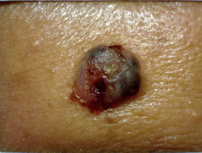
Nodulocystic basal cell carcinoma
A nodulocystic BCC is a small and slow-growing translucent lesion with rolled edges and telangiectasia.
- Nodular BCC is firm with a smooth surface.
- Cystic BCC is soft with jelly-like contents.
- A rodent ulcer has central ulceration.
Superficial basal cell carcinoma
Superficial BCC is the most common type of BCC found in younger adults.
- It is commonly found on the upper trunk and shoulders.
- It consists of a slightly scaly, irregular plaque with a thin, translucent rolled border.
- There may be multiple microerosions.
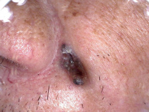
Morphoeic basal cell carcinomas
Morphoeic BCC is also known as morpheaform or sclerosing BCC.
- It is usually found in mid-facial sites.
- It consists of a waxy, scar-like plaque with indistinct borders and a wide and deep subclinical extension.
- It may infiltrate the cutaneous nerves (perineural spread).
Basosquamous basal cell carcinoma
Basosquamous BCC is also known as mixed BCC and SCC.
- It has an infiltrative growth pattern.
- It consists of a waxy, scar-like plaque with indistinct borders and a wide and deep subclinical extension.
- It is potentially more aggressive than other forms of BCC.
Adenoid basal cell carcinoma
Adenoid BCC is a rare histological variant of BCC.
- It consists of a lace-like arrangement of malignant cells around connective tissue.
- These are nodular or ulcerated lesions of a lower grade than nodular and morphoeic BCC.
- There is no predisposition for a particular body site [7].
Fibroepithelial tumour of Pinkus
Fibroepithelial tumour of Pinkus is a warty plaque, usually found on the trunk.
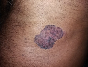
What are the clinical features of basal cell carcinoma in skin of colour?
The diagnosis of BCC in skin of colour can be difficult due to its rarity and pigmentation. More than 50% of BCCs found in skin of colour are pigmented with a ‘pearly’ brown or black appearance.
Nodular BCCs are the predominant subtype found in skin of colour, while morphoeic BCCs are uncommon. Adenoid BCC is more common than in Caucasians.
What are the complications of basal cell carcinoma?
Complications of BCCs may include:
- Recurrence or persistence after treatment
- New lesions
- Neglected advanced BCC
- Very rarely, metastatic BCC.
Recurrence of basal cell carcinoma
Basal cell carcinoma has an estimated 3–10% 5-year recurrence rate depending on whether the treated lesion is a primary BCC or is itself a recurrence. Most recurrences are evident 4–12 months after treatment. Recurrence is more likely with:
- An incomplete excision or narrow margins at the primary excision
- Morphoeic, micronodular, and infiltrative BCC subtypes
- BCCs located on the head and neck [8].
The type of treatment received can also influence recurrence rates. Higher recurrence rates (~7–10%) may be seen with all treatment methods other than Mohs micrographic surgery (~1%) [8].
Development of new basal cell carcinomas
There is a significantly increased risk of developing new BCCs after a previous BCC. Estimates from various studies have suggested a 3-year cumulative risk of between 30 and 77% for new BCC formation. Factors associated with new BCC formation may include:
- Older age
- Male sex
- Multiple non-melanoma skin cancers on initial presentation
- Truncal BCC [8].
Advanced basal cell carcinoma
Advanced BCCs are often a result of neglect as BCCs are very slow-growing tumours, often taking months to years to reach this stage. They may be several centimetres in diameter. Advanced BCCs may infiltrate tissues below the skin and are very difficult to treat surgically.
Metastatic basal cell carcinoma
Metastatic or invasive BCCs are rare, with an estimated occurrence rate of 0.03–0.55%. The primary BCC is often large, neglected or recurrent, and located on the head and neck region. It may arise in sites exposed to ionising radiation or ones that have had multiple prior treatments. Metastatic BCCs can be fatal.
How is basal cell carcinoma diagnosed?
Basal cell carcinomas are diagnosed by their typical appearance clinically and on examination using a dermatoscope [3]. The diagnosis is confirmed by incisional biopsy or excisional biopsy.
Most diagnoses occur at over 50 years of age and peak at around 70–80 years of age in both men and women, regardless of skin colour [5]. Men are much more likely to develop BCC — at double the incidence rate of women in some locations [5,8].
Staging basal cell carcinomas
In patients with advanced BCCs, it can be important to determine the depth of tumour invasion and the spread to surrounding tissue. The Union for International Cancer Control (UICC) 8th edition staging system for cutaneous carcinomas (skin cancers) is a commonly used system.
Tumour staging for cutaneous carcinomas
TX: Primary tumour cannot be assessed
T0: No evidence of primary tumour
Tis: Carcinoma in situ
T1: Tumour ≤ 2 cm, without high-risk features
T2: Tumour > 2 and < 4 cm
T3: Tumour with the invasion of maxilla, mandible, orbit or temporal bone
T4a: Tumour with gross cortical bone/marrow invasion
T4b: Tumour with skull base or axial skeleton invasion, including foraminal involvement and vertebral foramen involvement to the epidural space
Nodal staging for cutaneous carcinomas (non-head and neck)
NX: Regional lymph nodes cannot be assessed
N0: No regional lymph node metastasis
N1: Metastasis in one local lymph node ≤ 3 cm
N2: Metastasis in a single ipsilateral lymph node > 3 cm and ≤ 6 cm or in multiple ipsilateral nodes ≤ 6 cm.
N3: Metastasis in lymph node ≥ 6 cm
What is the differential diagnosis of basal cell carcinoma?
There are several lesions that can be mistaken for a basal cell carcinoma, especially:
- Actinic keratosis
- Squamous cell carcinoma (SCC)
- Intraepidermal SSC
- Seborrhoeic keratosis
- Dermatofibroma
- Intradermal naevus (mole)
- Sebaceous hyperplasia
- Melanocytic naevus
- Pyogenic granuloma
- Trauma
- Microcystic adnexal carcinoma
- Cutaneous T-cell lymphoma
- Merkel cell carcinoma
- Trichoepithelioma/trichoblastoma/trichoadenoma
- Atypical fibroxanthoma
- Metastasis to skin.
Malignant lesions, such as cutaneous SCC and melanoma, have a peak incidence at the same approximate age (> 50 years) as BCC and may be present simultaneously.
What is the treatment for basal cell carcinoma?
Treatment options are the same, regardless of skin colour. Excisional biopsy with a 3–5 mm margin is the standard and recommended treatment for most BCCs.
Smaller margins increase the risk of recurrence. Deeper incisions and more complex wound closure (flap or skin graft) may be required for larger BCCs. The removed specimen is then examined by a pathologist for a final diagnosis and disease staging [9].
Potential side effects of treatment can include infection, scarring, and temporary or permanent loss of sensation due to nerve severing.
Cryotherapy
Cryotherapy is the treatment of a superficial skin lesion by freezing it, usually with liquid nitrogen [9]. It is suitable for small superficial BCCs (≤ 1cm) on covered areas of trunk and limbs, and not advised to treat BCCs on head, neck and knees. Cryotherapy:
- Consists of a double freeze-thaw application technique.
- Results in a blister that crusts over and heals within several weeks.
- May require wound dressing if the wound is at risk of rubbing.
Complications and side effects of cryotherapy include:
- The procedure leaving a permanent white mark.
- When applied to the back of a hand, there is a risk of damaging the underlying tendons
- Application over a nerve involved in sensation may result in temporary numbness for weeks or months.
Shave, curettage and electrocautery
Shave, curettage, and electrocautery are superficial forms of surgery. These techniques require local anaesthesia but are quick to perform and do not require sutures [9].
- These techniques are useful for small, well-defined nodular or superficial BCCs.
- Suitable lesions are usually located on the trunk or limbs.
- The wound is left open to heal by secondary intention healing (it is left to heal by itself).
- Moist wound dressings lead to healing within a few weeks.
- The quality of the eventual scar is variable.
Mohs margin-controlled micrographic excision
Mohs micrographic controlled surgery involves removing skin tissue in successively deeper horizontal sections under a local anaesthetic. Each section is examined with a microscope to identify cancerous tissue. Sections are continuously removed until no further cancerous tissue is identified [10].
- Mohs margin-controlled micrographic excision is used in high–risk areas of the face, such as around eyes, lips, and nose.
- Suitable for ill-defined, morphoeic, infiltrative and recurrent subtypes.
- Large defects are repaired by flap or skin graft.
Mohs margin-controlled micrographic excision has very high cure rates when performed by trained dermatologists or dermatological surgeons.
Imiquimod cream
Imiquimod is a topical immune response-modifying drug [9,10]. Imiquimod cream is used when surgical removal is inappropriate.
- Imiquimod is best used for superficial BCCs < 2 cm in diameter.
- It is applied 3–5 times each week, for 6–16 weeks.
- Imiquimod results in a variable inflammatory reaction, that is greatest at three weeks.
Side effects and complications of imiquimod cream include:
- Minor scarring
- Pain or itching
- An inflammatory reaction, maximal 3–4 days after the use of the cream
- Ulceration.
Fluorouracil cream
5-Fluorouracil cream is a topical cytotoxic agent [9,10]. It is used to treat small superficial BCCs.
- 5-Fluorouracil requires a prolonged course (eg, twice daily for 6–12 weeks).
- It causes an inflammatory reaction.
- There are high recurrence rates with the use of 5-fluorouracil.
Side effects and complications with the use of 5–fluorouracil cream include:
- Inflammation and photosensitivity over the applied area (this is expected).
- Significant pain
- Erythema multiforme
- Permanent hyperpigmentation
- Scarring.
Photodynamic therapy
Photodynamic therapy (PDT) refers to a technique in which BCC is treated with a photosensitising chemical and exposed to light several hours later [9,10].
- Topical photosensitisers used in PDT include aminolevulinic acid lotion and methyl aminolevulinate cream.
- Photodynamic therapy is suitable for low-risk small, superficial BCCs and thin nodular BCCs or in patients with multiple BCCs, and best avoided if tumours are sites where they are at high risk of recurrence.
- Best avoided if the tumour in a site at high risk of recurrence.
- It results in an inflammatory reaction that is greatest 3–4 days after the PDT.
- Treatment is repeated seven days after the initial treatment.
- There are excellent cosmetic results.
Side effects and complications of photodynamic therapy include:
- Pain or itching
- An inflammatory reaction, maximal 3–4 days after the procedure.
Radiotherapy
Radiotherapy or X-ray treatment can be used to treat primary BCCs or as adjunctive treatment, if margins are incomplete [9,10]. Radiation is mainly used if surgery is not suitable. It is best avoided in young patients and in people with genetic conditions that predispose them to skin cancer. The best cosmetic results are achieved using multiple fractions.
Side effects and complications with the use of PDT include:
- Inflammation and scarring of the treated area is expected
- Late development of new BCCs, or BCC recurrence
- Radiodermatitis.
What is the treatment for advanced or metastatic basal cell carcinoma?
Advanced or metastatic BCCs require multidisciplinary consultation. A combination of treatments may be used including:
- Surgery
- Radiotherapy
- Chemotherapy
- Targeted cancer therapy using hedgehog signalling pathway inhibitors, such as vismodegib.
What is the recommended follow-up for basal cell carcinoma?
There are no established guidelines for the follow-up after the removal of a BCC. An annual skin check is recommended for anyone who has had a BCC, given the estimated 30–77% cumulative risk of experiencing another one within three years. [8–10].
Year-round protection against excessive sun exposure is essential in all skin types.
- Stay indoors or under the shade in the middle of the day.
- Wear covering or sun-protective clothing.
- Apply high protection factor SPF50+ broad-spectrum sunscreens generously to exposed skin if outdoors.
- Avoiding indoor tanning (sun beds and solaria).
What is the outcome for basal cell carcinoma in skin of colour?
Basal cell carcinomas are very slow-growing tumours and are unlikely to reach an advanced stage unless significantly neglected. As long as the tumour has been completely excised, BCC does not recur. However, another may develop elsewhere.
The prognosis is of the rare metastatic BCC is very poor, with a median survival time of 8 months following diagnosis [3,10].