What is a blue naevus?
A blue naevus is a common type of melanocytic naevus in which the pigment is situated deep in the dermis. It is also known as a blue neuronaevus and a dermal melanocytoma.
What are the clinical features of a blue naevus?
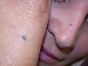
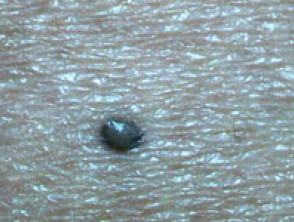
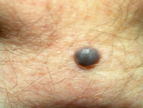
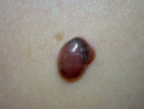
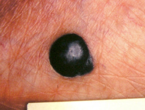
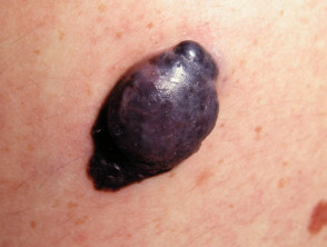
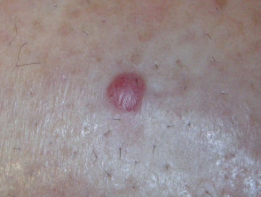
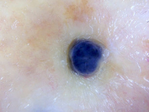
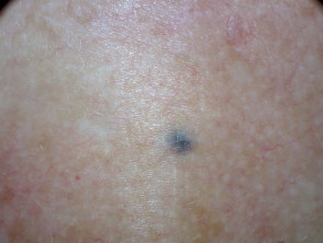
A blue naevus is a well-circumscribed round or oval macule, papule or nodule of a uniform steel blue colour. The border fades gradually into the surrounding skin.
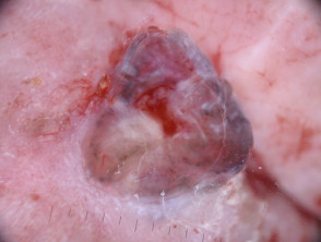
- The common blue naevus is usually 0.5–1 cm in diameter. The cellular blue naevus is more nodular and is at least 1 cm in diameter.
- The colour of blue naevi can also vary, usually being composed of blue to grey hues, but they are sometimes brown or yellowish.
- Blue naevi are usually found on the distal extremities (on the dorsum of the hands and feet), buttocks, scalp and face although they can occur anywhere on the body. They have rarely been reported in the vagina, the spermatic cord, the lymph nodes, the uterine cervix, the prostate and the oral mucosa.
In adults, the lesion is often long-standing. The most common age of onset is late childhood or adolescence.
Blue naevi
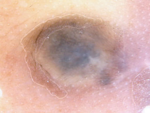
What are the dermoscopic features of a blue naevus?
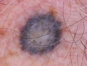
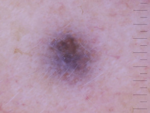
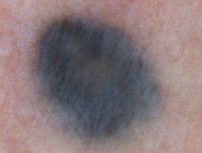
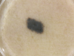
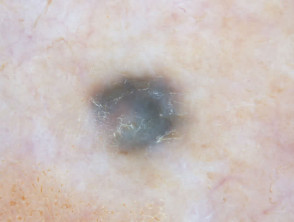
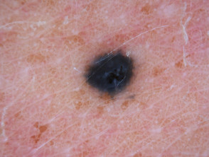
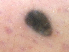
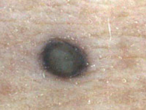
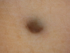
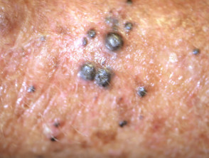
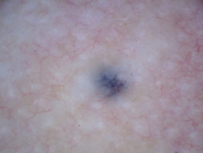
The dermoscopic hallmark of a blue naevus is homogeneous blue pigmentation. In some exceptional cases, they may exhibit blue globules and dots [1].
Dermoscopic features of a blue naevus are:
- Structureless pattern
- Uniform blue colour
- Lack of any other structures
- A well-defined border that fades into the outer skin.
- A diffuse blue-white veil appearance over the entire surface.
The presence of any of the following structures should warrant further investigations for melanoma: network, dots, clods, streaks, vessels, additional colours.
Dermoscopic images of blue naevi
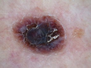
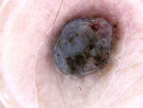
What is the dermoscopic differential diagnosis of a blue naevus?
The primary concern of a lesion presenting as a blue naevus with unknown history is that it could be one of the following:
- Nodular melanoma
- Melanoma metastasis
- Blue tattoo (eg, to indicate the site of radiation).
Differential diagnosis of blue naevus
What is the histological explanation of features of a blue naevus?
The histological explanation of a blue naevus is:
- Blue colour: pigment deep in the reticular dermis
- Structureless: the heavily pigmented dendritic melanocytes are randomly arranged with no relationship to the architecture of the overlying epidermis and so appear structureless on dermoscopy.
- Blue-white veil: orthokeratotic surface.