Introduction
Branchial cleft cysts are remnants of embryonic development and result from a failure of obliteration of one of the branchial clefts, which in fish develop into gills.
Histology of branchial cleft cyst
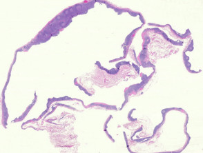
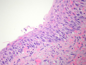
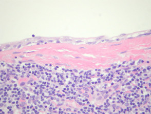
A branchial cleft cyst is often surrounded by lymphoid tissue (figure 1). The lining of the cyst is usually a stratified squamous epithelium (figure 2). The lining may also be a columnar ciliated epithelium. Often there are marked inflammatory changes and the epithelium overlying the lymphoid tissue is attenuated/absent (figure 3). Smooth muscle is rarely seen in the wall. Mucous glands and cartilage may also sometimes be seen in the wall.
Branchial cleft cyst pathology
Special studies for branchial cleft cyst
None are needed.
Differential diagnosis of branchial cleft cyst pathology
Cutaneous ciliated cyst – Smooth muscle, mucous glands and cartilage are not seen in the walls of cutaneous ciliated cysts.
Metastatic squamous cell carcinoma – Squamous cell carcinoma metastatic to neck lymph nodes can be extremely difficult to distinguish from branchial cleft cysts, particularly when the metastatic tumour is cystic.
Bronchogenic cyst – Usually arise in the midline, almost always have smooth muscle or glands in the wall and are generally not associated with lymphoid tissue.