Introduction
Calcifying aponeurotic fibromas present at a young age on the hands and feet, as small nodular or infiltrative masses.
Histology of calcifying aponeurotic fibroma
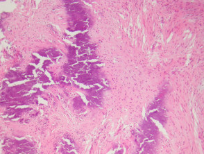
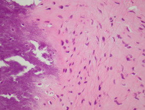
In calcifying aponeurotic fibroma, sections show an irregularly shaped dense mass of fibrous tissue with foci of calcification (figures 1, 2). The fibrous tissue is quite cellular and appears to infiltrate the surrounding adipose tissue (figure 1).
Rarely extramedullary haematopoiesis may be seen.
Calcifying aponeurotic fibroma pathology
Special studies for calcifying aponeurotic fibroma
None are generally needed.
Differential diagnosis of calcifying aponeurotic fibroma pathology
Plantar/palmar fibromatosis – Fibromatosis usually occurs later in life. Also, the mass is usually well defined as opposed to the infiltrative border of calcifying aponeurotic fibroma.