Cutaneous carcinoid tumours represent metastases from primaries in the lung or gastrointestinal system.
Cutaneous features of carcinoid syndrome including flushing, rosacea, scleroderma and pellagra.
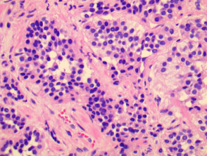
Histology of carcinoid
Microscopically tumours are composed of solid islands and nests of uniform cells. Thin collagenous septa extend between the tumour nests.
Gastric carcinoid pathology
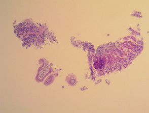
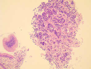
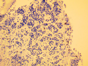
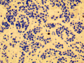
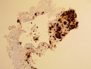
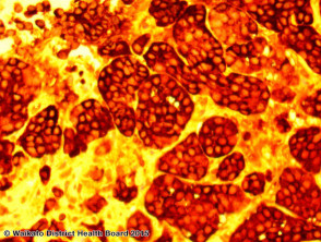
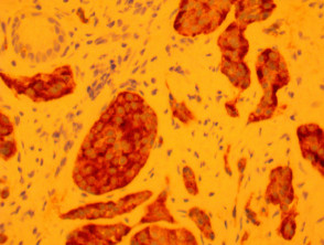
Images provided by Dr Duncan Lamont, Waikato Hospital
Special studies in carcinoid
Tumour cells are positive for neuron-specific enolase, chromogranin and synaptophysin.
On electron microscopy dense-core granules can be seen in the cytoplasm.
Differential diagnosis of carcinoid
Primary adnexal poroid neoplasm
Hailey-Hailey disease