Introduction
Clear cell mesenchymal neoplasm (CCMN, also called distinctive clear cell mesenchymal neoplasm) is a rare dermal tumour of uncertain lineage. The tumours have a benign clinical course but histologically closely resemble malignant tumours including dermal metastases.
Histology of clear cell mesenchymal neoplasm
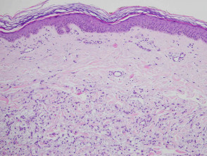
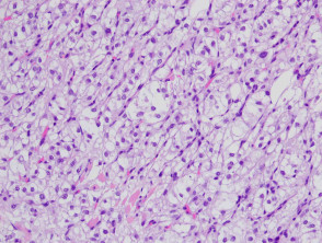
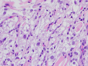
Histologically, these tumours arise in the dermis and are usually well circumscribed (figure 1). The cells of a tumour are clear, large cells (approximately 6–8 times the size of a stromal lymphocyte and about half the size of a subcutaneous adipocyte) that are oval to polygonal in shape with abundant clear cytoplasm (figure 2). Closer inspection reveals that, while many of the cells are optically clear, others contain lacy, reticular cytoplasm that has a slightly granular appearance (figure 3).
Clear cell mesenchymal neoplasm pathology
Special stains for clear cell mesenchymal neoplasm
These tumours are negative for immunohistochemical markers for various keratins, melanoma markers, and markers often seen in renal cell carcinoma (Pax-8, RCC).
Differential diagnosis of clear cell mesenchymal neoplasm
Metastatic clear cell carcinoma including renal cell carcinoma: This is probably the most important differential diagnosis. Numerous immunohistochemical studies including PAX-8, AE1/3, RCC, EMA need to be confirmed to be negative to exclude this possibility
Melanoma — CCMN is negative with the immunohistochemical markers for melanoma
PEComa — This tumour usually expresses melanocytic markers