Introduction
Buy a dermatoscope from DermNet's Amazon store
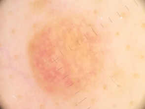
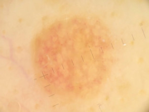
This page includes galleries of images of skin lesions grouped by the lesion diagnosis and by the camera/dermatoscope used to capture them.
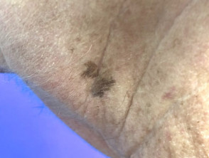
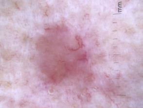
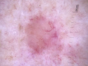
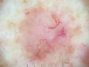
Images were taken of melanocytic naevi (3), solar lentigines (3), seborrhoeic keratosis, basal cell carcinoma (3), squamous cell carcinoma in situ (2), and superficial spreading melanoma in situ using various cameras and dermatoscopes for comparative purposes.
Not all images are displayed due to patient, lesion, or image-quality issues.
Devices used were:
- iPhone X with Dermlite DL4, DL3, Hybrid, HÜD, GL; Illuco; Opticlar;
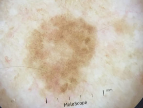
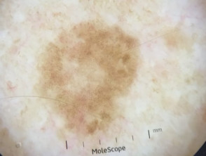
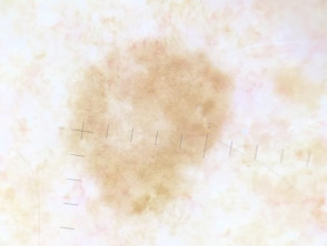
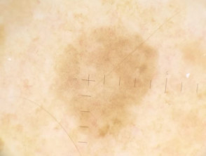
- iPhone 6 with Heine iC1 and Molescope
- iPod Touch 5 with Fotofinder Handyscope
Polarised dermoscopy views were taken with Dermlite DL4, DL3, Hybrid, Hüd, GL, Illuco, Opticlar, iC1, Handyscope
Unpolarised dermoscopy views were taken with Dermlite DL4, DL3, Hybrid, Illuco, Opticlar, iC1, Handyscope
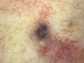
Benign lesions
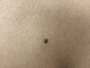
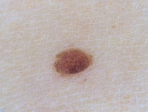
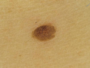
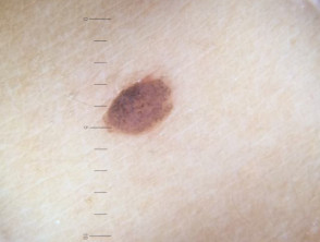
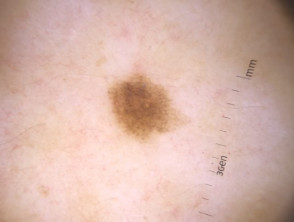
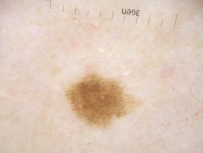
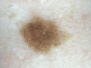
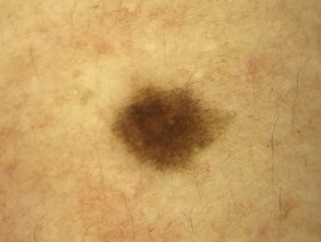
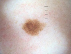
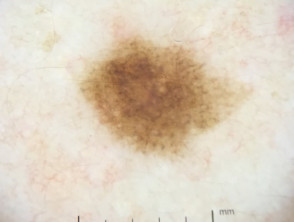
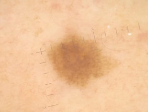
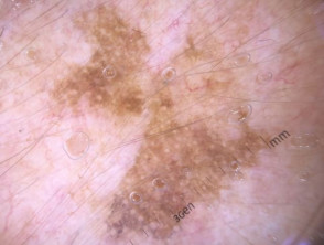
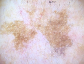
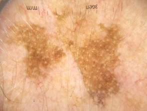
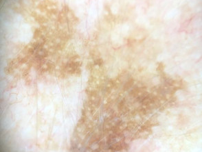
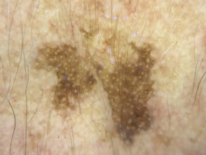
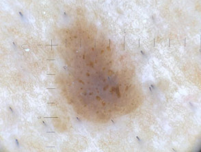
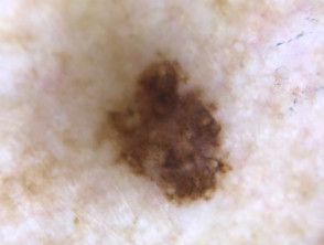
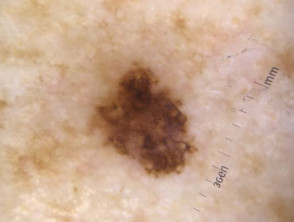
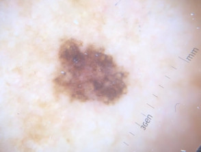
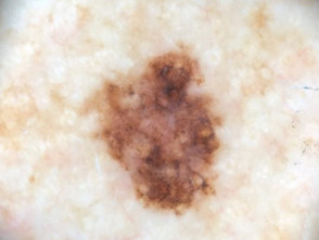
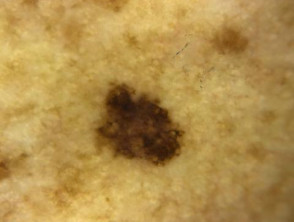
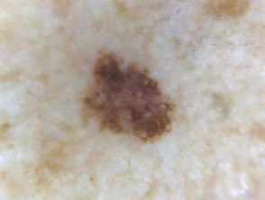
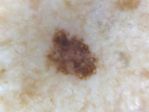
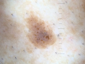
Dermoscopic images of melanocytic naevi
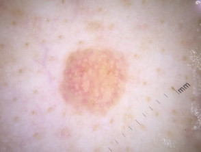
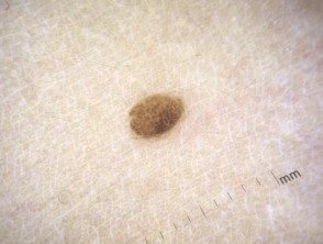
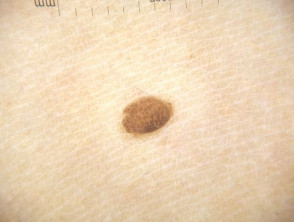
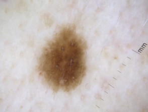
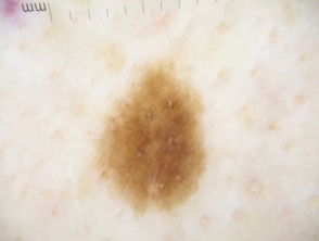
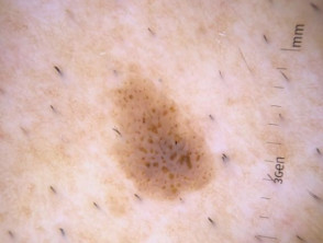
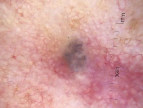
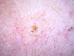
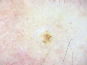
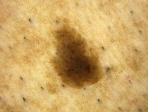
Dermal naevus example 1

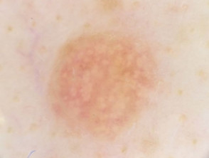
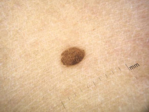
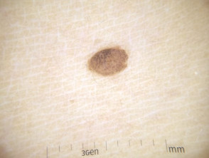
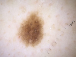
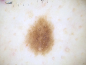
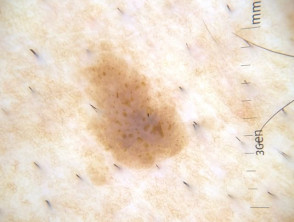
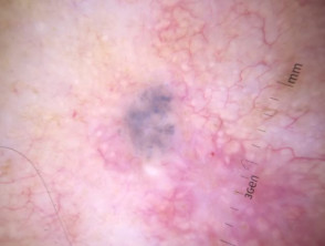
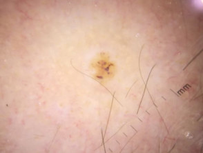
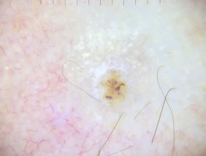
Melanocytic naevus example 2
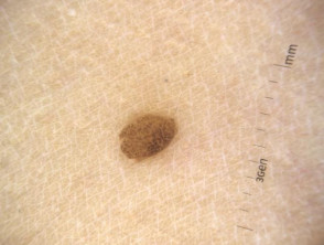
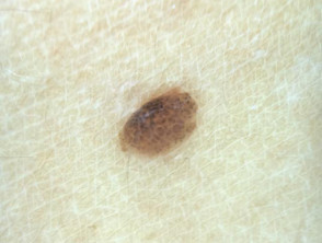
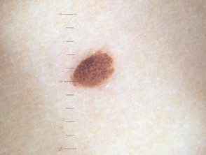
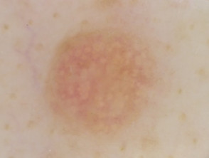
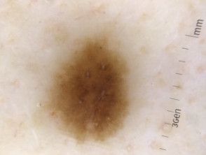
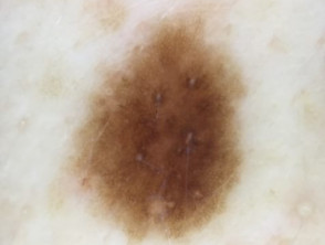
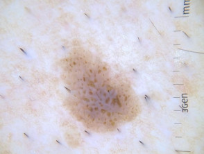
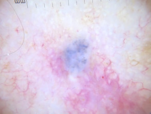
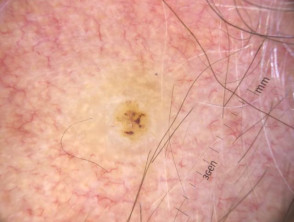
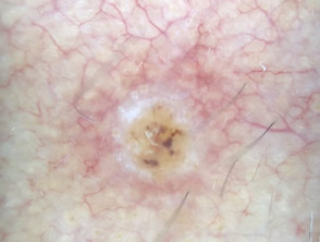
Melanocytic naevus example 3
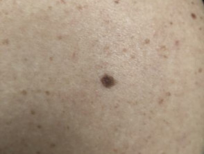
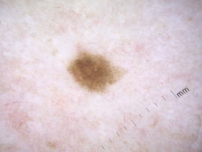
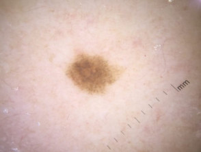
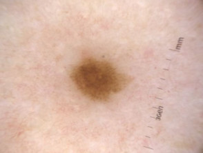
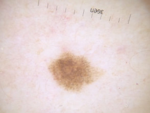
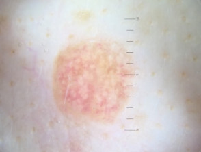
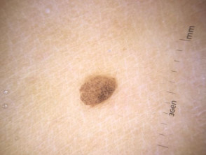
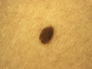
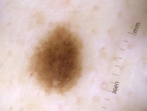
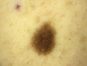
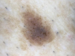
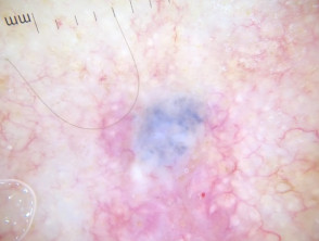
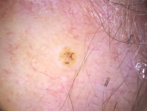
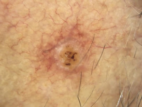
Melanocytic naevus example 4

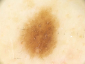
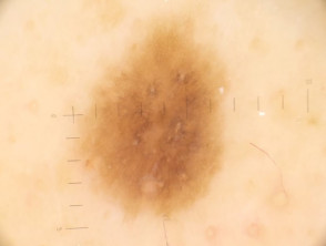
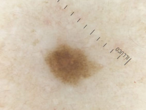
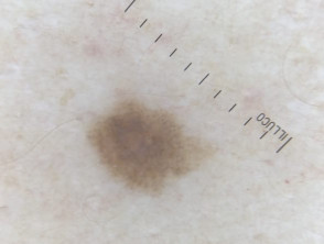
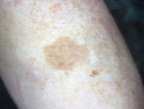
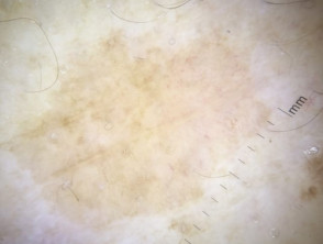
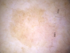
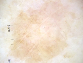
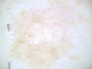
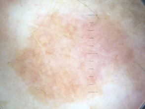
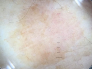
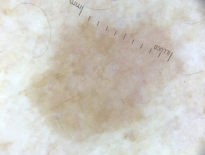
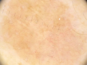
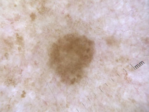
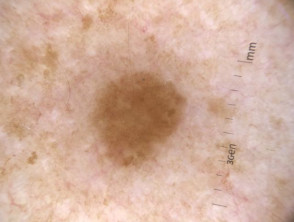
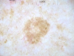
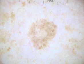
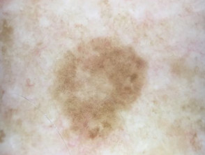
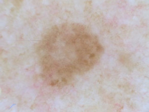
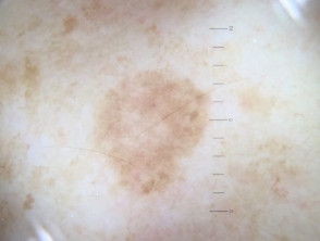
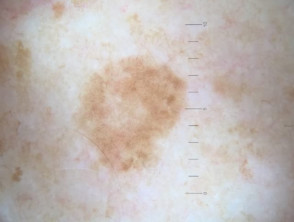
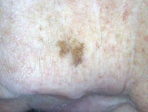
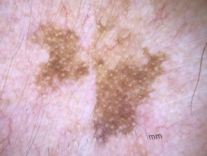
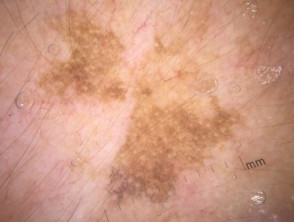
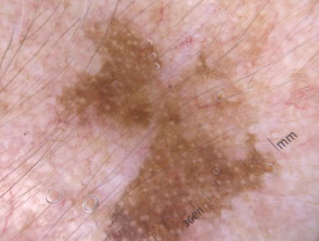
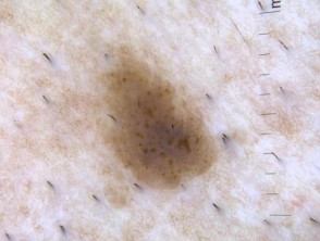
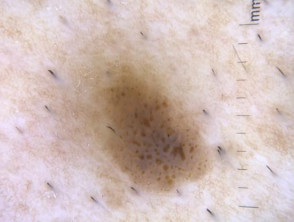
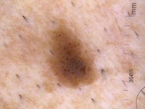
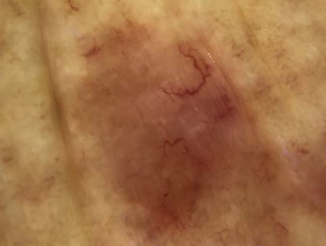
Dermoscopic images of solar lentigines
Solar lentigo example 1
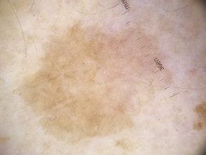
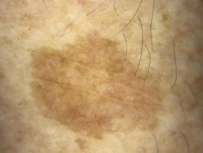
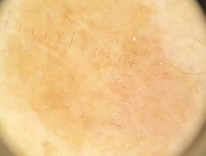
Solar lentigo example 2

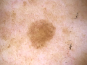
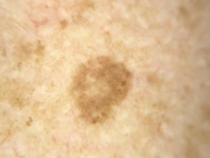

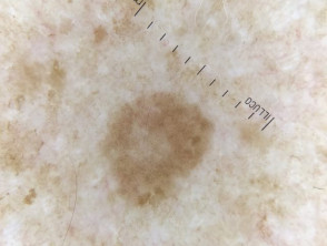
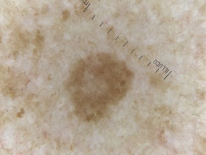
Solar lentigo example 3
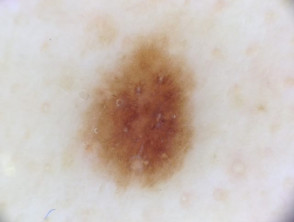
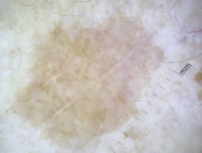
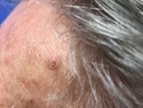
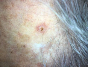
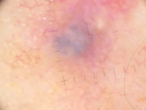
Dermoscopic images of seborrhoeic keratoses
Seborrhoeic keratosis


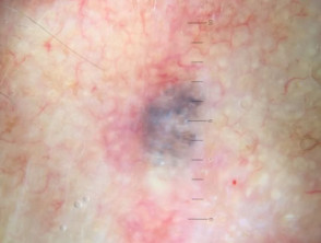
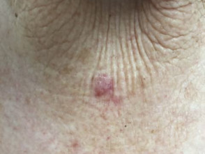
Malignant lesions
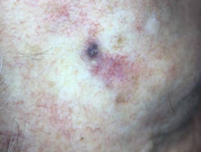
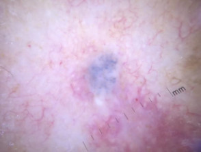
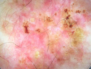
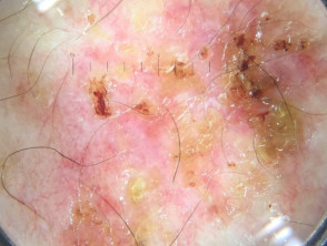
Dermoscopic images of basal cell carcinoma
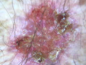
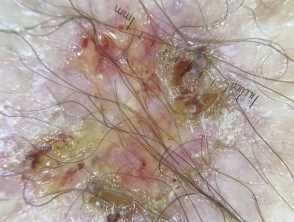
Basal cell carcinoma example 1
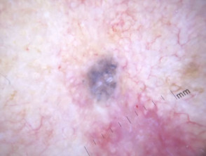
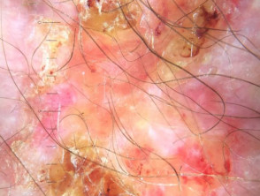
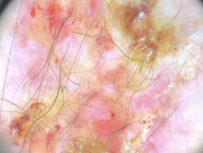
Basal cell carcinoma example 2
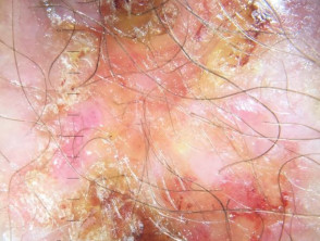
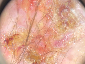
Basal cell carcinoma example 3
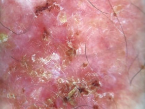
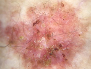
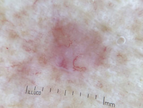
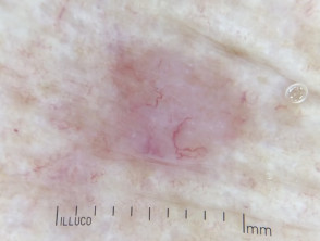
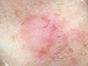
Dermoscopic images of squamous cell carcinoma in situ
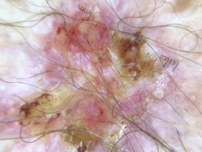
Squamous cell carcinoma in situ example 1
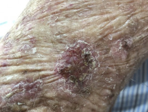
Squamous cell carcinoma in situ example 2
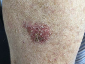
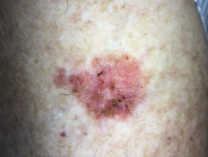
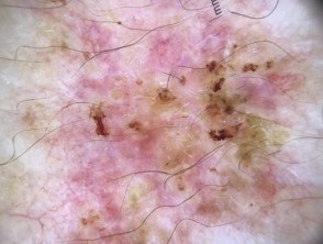
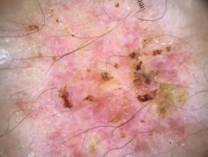
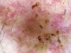
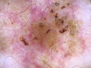
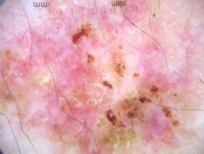
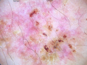

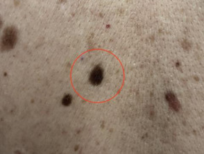
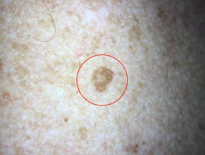
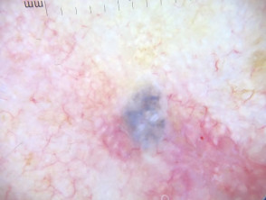
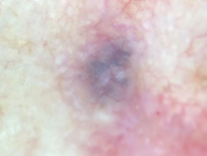
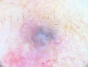
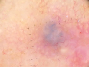
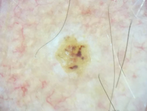
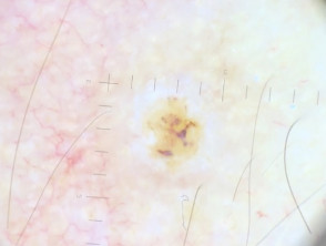
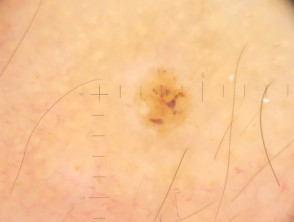
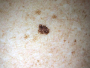
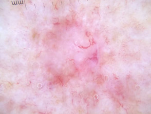
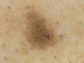
Dermoscopic images of superficial spreading melanoma in situ
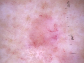
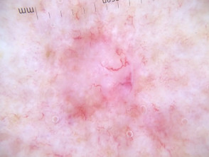
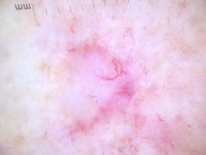
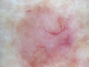
Superficial spreading melanoma in situ
Dermatoscopes
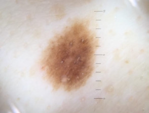
Dermoscopic images taken with Dermlite DL4 (iPhone X)
Dermlite DL4
Dermoscopic images taken with Dermlite DL3 (iPhone X)
Dermlite DL3
Dermoscopic images taken with Dermlite Hybrid (iPhone X)
Dermlite Hybrid
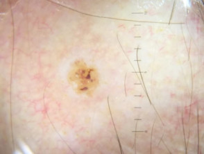
Dermoscopic images taken with Dermlite Hüd (iPhone X)
Dermlite HÜD
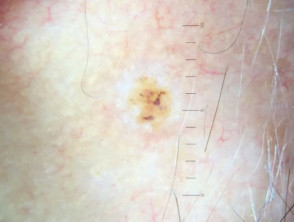
Dermoscopic and macro images taken withDermlite GL (iPhone X)
Dermlite GL
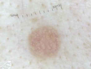
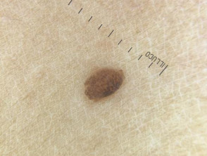
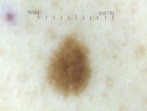
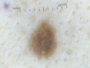
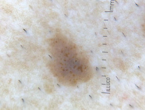
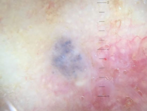
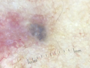
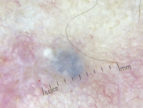
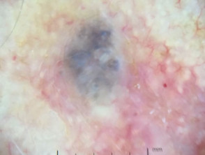
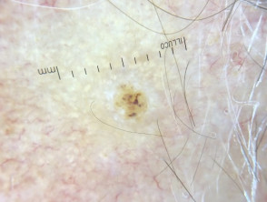
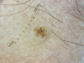
Dermoscopic images taken with Illuco (iPhone X)
Illuco IDS 1100
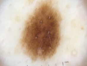
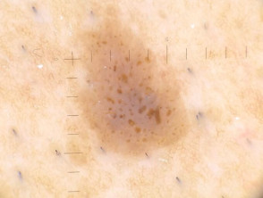
Dermoscopic images taken with Opticlar D-scope Compact (iPhone X)
Opticlar D-scope Compact Dermatoscope
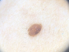
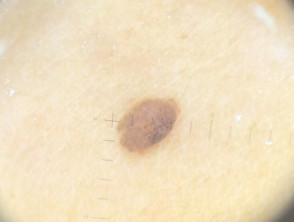
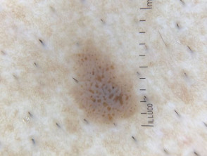
Dermoscopic images taken with Heine iC1 (iPhone 6)
Heine iC1

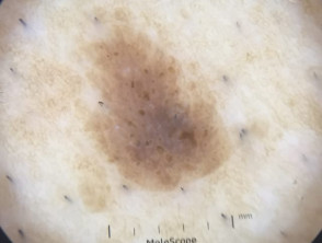
Dermoscopic images taken with Fotofinder Handyscope (iPod Touch)
Fotofinder Handyscope

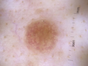
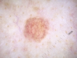
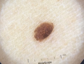
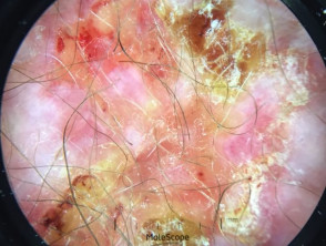
Dermoscopic images taken with MetaOptima Molescope II
Molescope II