This lesion falls within the group of benign follicular tumours.
Histology of dilated pore of Winer
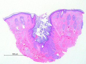
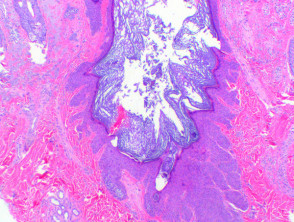
Scanning power view of dilated pore of Winer demonstrates a small invaginating epidermal process (Figure 1). This is comprised of a widened follicular infundibulum filled with a small amount of keratinous material (Figure 2). The infundibular epithelium is hyperplastic, forming small radiating strands at the base and sides of the pore (Figure 2).
Dilated pore of Winer pathology
Differential diagnosis of dilated pore of Winer
Pilar sheath acanthoma: In this lesion there is more prominent hyperplasia of the outer root sheath forming tumour nodules which push into the surrounding dermis. This tumour arises from the follicular isthmus where the corneocytes take on a more red-pink colour.
Trichofolliculoma: Radiating hair follicles in variable states of maturity are seen to extend from the central dilated pore or cystic structure.
Hair cortex comedo : This lesion demonstrates infundibular epithelium associated with a dense cornified plug with compact laminated corneocytes, and matrical and supramatrical epithelium representing attempts to form the hair cortex and shaft.