Introduction
This distinctive tumour is an adnexal tumour argued to arise from the apocrine or eccrine secretory coil or coiled duct.
Histology of eccrine spiradenoma
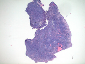
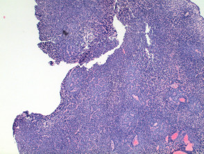
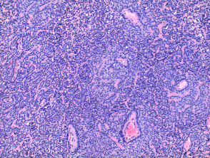
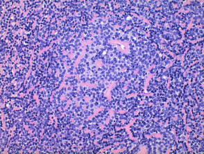
Low power view of eccrine spiradenoma shows a well-circumscribed tumour nodule arising within the dermis or superficial subcutis. The tumour is comprised of a diffuse dense basophilic cellular proliferation (Figures 1 and 2). In some cases a prominent vascular component can be seen (Figures 2 and 3). Eosinophilic hyaline deposits are seen in amongst the tumour cells as droplets and bands (Figure 4). A lymphocytic infiltrate is seen and when heavy can mimic lymphoid tissue.
Eccrine spiradenoma pathology
Variants of eccrine spiradenoma pathology
Brooke-Spiegler syndrome — no differences are seen between sporadic eccrine spiradenoma or those occurring in the context of this syndrome. Tumours with overlap features with eccrine spiradenoma can be seen.
Differential diagnosis of eccrine spiradenoma pathology
Cylindroma — in this tumour the low power view shows discrete polygonal tumour islands. There is frequently a more prominent population of S100 positive dendritic cells while in eccrine spiradenoma there is a lymphocytic infiltrate.
Spiradenocylindroma — some tumours features of both cylindroma and eccrine spiradenoma can be seen and are designated as overlap tumours.
Cutaneous lymphadenoma — in this tumour there is a lobular tumour of basaloid cells which may show peripheral palisading, and a dense mixed inflammatory cell infiltrate though predominantly lymphocytic.