What is epulis?
Epulis literally means of the gums and is a non-specific term used for tumours and tumour-like masses of the gingiva (gums). There are several lesions or growths of the gums that are referred to as epulis, however their clinical characteristics are unique and they should be discussed as single disease entities of their own.
| Epulis type | Description |
|---|---|
| Epulis fissuratum | Overgrowth of fibrous connective tissue around the edges of ill-fitting dentures |
| Giant-cell epulis | A peripheral giant cell granuloma that arises exclusively from the periodontal ligament enclosing the root of a tooth |
| Congenital epulis | Rare tumour of the newborn that arises from the mucosa of the gingiva |
Epulis fissuratum
Why does epulis fissuratum occur?
Epulis fissuratum is also referred to as inflammatory fibrous hyperplasia, denture epulis and denture-induced fibrous hyperplasia. The fibrous overgrowth is caused by chronic irritation of the denture flange (edge) against the area where the gums meet the inner cheek (alveolar vestibular mucosa). Because bone under the denture is constantly changing due to bone loss, the bony support for the denture base becomes unstable which results in ill-fitting dentures and epulis fissuratum.
Who get epulis fissuratum?
The condition is most common in the elderly as the need for dentures increases with age. However, it is becoming less of a problem nowadays as dental technology to maintain and restore teeth is much more advanced. The condition appears to be more common in women than men.
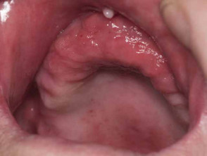
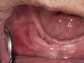
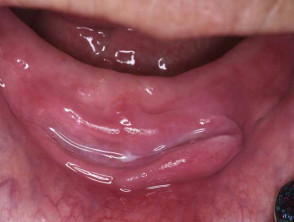
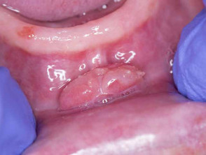
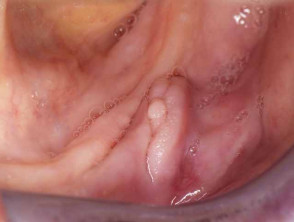
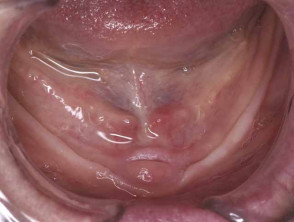
What are the signs and symptoms?
The lesions made up of excess tissue are usually firm, fibrous and pink. One part of the lesion is found under the denture while the rest protrudes into the cheek area. The internal and external parts of the lesion are separated by a deep groove in which the denture flange sits. Sometimes the irritation can be severe enough to cause redness and ulceration, particularly at the bottom of the groove where the denture rests.
How do you treat epulis fissuratum?
The lesions of epulis fissuratum can be surgically cut out. Although it is very uncommon for these lesions to be associated with oral squamous cell carcinoma, as a precautionary measure the removed lesion should be sent for microscopic testing.
In addition to removing lesions, dentures should be re-modelled to fit properly into the gums to prevent further irritation and development of epulis fissuratum.
Giant cell epulis
Why does giant cell epulis occur?
Giant cell epulis is also referred to as peripheral giant cell granuloma, giant cell reparative granuloma, osteoclastoma and myeloid epulis. It is not clear why lesions of giant cell epulis occur but it may be a response to injury. In addition, many cases have cells expressing surface receptors for oestrogen, which has led to speculation that hormonal influences may play a role in their development.
Who gets giant cell epulis?
Giant cell epulis may occur at any time, although it appears to be most commonly diagnosed between 40-60 years of age. In adults it occurs mainly in women.
What are the signs and symptoms?
Lesions are often fibrous and present as a gingival lump emerging between two teeth. The lesion grows quickly and may exceed 4cm in diameter, but most remain less than 2cm. It can grow into an irregular red fleshy mass that may ulcerate or bleed. Sometimes the giant cell epulis may invade underlying bone.
How do you treat giant cell epulis?
Treatment involves surgical excision of the lesion and curettage of any underlying bony defect. The affected teeth may also need to be extracted or scaling and root planing performed. A recurrence rate of 10% or more has been reported and re-excision may be required.
Congenital epulis
Why does congenital epulis occur?
Congenital epulis is also referred to as congenital granular cell myoblastoma, granular cell epulis of infancy, and granular cell fibroblastoma. It is not clear why congenital epulis occur but they are thought to originate from primitive mesenchymal cells of neural crest origin.
Who gets congenital epulis?
This is a rare congenital condition that appears at birth. It occurs much more commonly in female than males with a ratio of 8:1.
What are the signs and symptoms?
Infants are born usually with a mass protruding out their mouth. The lesion is almost exclusively found on the anterior alveolar ridges of the newborn. In 10% of cases multiple lesions may be present. Lesions may range from 0.5 to 2cm in size, and in rare cases reach up to as much as 9cm. The lesion is a soft, pedunculated and sometime lobulated nodule of the alveolar mucosa.
If the epulis is too large it may interfere with breathing and feeding.
How do you treat congenital epulis?
Most epulis tend to spontaneously regress and disappear over the first 8 months of life. Hence, if the lesion is small there may be no need for treatment. Larger lesions that may interfere with breathing and/or feeding may need to be surgically removed. Carbon dioxide laser has been used successfully to remove large lesions. Recurrences of epulis have not been reported and residual remnants appear not to interfere with tooth eruption.