Introduction
Also known as florid nipple papillomatosis and papillary adenoma, these lesions present as nipple erythema or nodules which may mimic mammary Paget disease.
Histology of erosive papillomatosis of the nipple
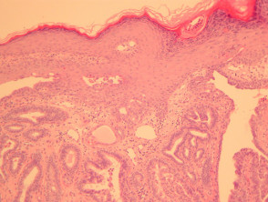
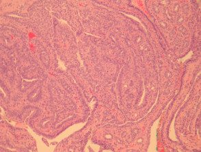
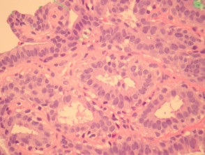
In erosive papillomatosis of the nipple, sections show a papillary epithelial proliferation which connects with the overlying epidermis (figure 1). The lesion consists of fibrovascular cores lined by bland cells (figure 2). The cells are usually at least 2 layers thick and consist of a myoepithelial layer and an epithelial layer which may exhibit apocrine differentiation (figure 3)
Erosive papillomatosis of the nipple pathology
Special studies of erosive papillomatosis of the nipple
None are generally needed.
Differential diagnosis of erosive papillomatosis of the nipple
Carcinoma – Breast carcinoma invading the nipple will show more nuclear atypia, usually lack a papillary growth pattern and lacks the dual myoepithelial/epithelial lining.