Erythema gyratum repens is a clinically distinctive figurate or annular erythema that is usually associated with underlying malignancy. The eruption is rapidly migrating and composed of concentric rings forming a wood-grain pattern.
Histology of erythema gyratum repens
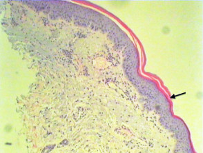
Histological features of erythema gyratum repens are not diagnostic and include hyperkeratosis, parakeratosis, acanthosis and spongiosis. There may be perivascular lymphohistiocytic infiltrate in the papillary dermis (Figure 1, arrow shows parakeratosis).
Erythema gyratum repens pathology
Differential diagnosis of erythema gyratum repens
Histological differential diagnosis is broad as features are quite nonspecific. Fortunately the clinical appearance is distinctive. Biopsy may be undertaken to exclude clinical differentials including erythrokeratoderma variabilis, resolving pityriasis rubra pilaris, bullous pemphigoid and epidermolysis bullosa aquisita.