Introduction
Inflammatory Linear Verrucous Epidermal Naevus (ILVEN) is a distinct form of inflammatory epidermal naevus with a characteristic histopathology. Rarely, there may be associated abnormalities involving other ectodermal structures.
Histology of inflammatory linear verrucous epidermal naevus
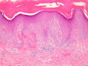
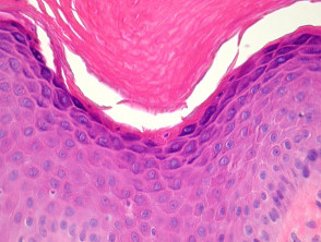
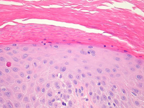
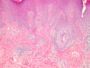
In ILVEN, sections show skin with psoriasiform epidermal hyperplasia with alternating orthokeratosis and parakeratosis (figure 1). The orthokeratotic areas overlie depressed epidermis with marked hypergranulosis (figure 2). The zones of parakeratosis overlie hypogranulosis (figure 3). There are spongiotic epidermal changes and small microvesicles. There is a mixed dermal inflammatory response (figure 4).
Inflammatory Linear Verrucous Epidermal Naevus pathology
Special studies for inflammatory linear verrucous epidermal naevus
None are generally needed.
Differential diagnosis of inflammatory linear verrucous epidermal naevus
Epidermal naevus – This resembles seborrheic keratosis and generally lacks the inflammatory changes and characteristic changes in the stratum corneum seen in ILVEN.
Psoriasis — Distinction can be difficult without clinical correlation. Neutrophilic pustulation is generally not a feature of ILVEN.