What are keloids and hypertrophic scars?
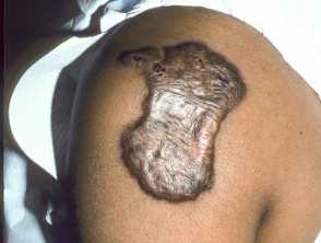
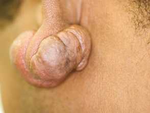
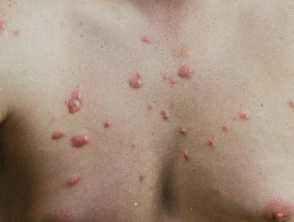
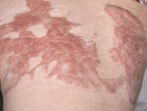
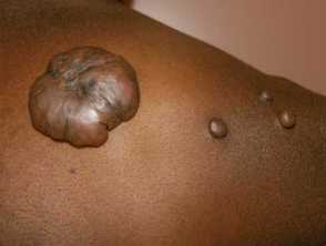
A keloid scar is a firm, smooth, hard growth that occurs as a result of excessive scar formation. Keloids occur after skin injury; rarely, keloids can occur spontaneously without any significant preceding skin injury. They may develop on any part of the body and extend beyond the original wound margin, although the upper chest, shoulders, ears, and neck are especially prone to them.
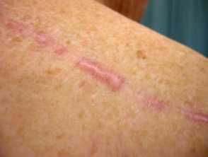
Unlike keloid scars, hypertrophic scars are limited to the area of damaged skin. They are prone to occur when there is a lot of tension on a healing wound, the resultant scar is thicker than usual. Hypertrophic scars are more likely to regress and resolve compared to keloids as these tend to persist.
Keloid and hypertrophic scar
Who gets keloids and hypertrophic scars?
Hypertrophic scarring is common and can occur in all races and ages whereas keloid scars are less common and are more frequent in those with Fitzpatrick skin types III to VI.
Keloids are self-reported in 16% of Black individuals, and Chinese individuals are more likely to develop them when compared with those of Indian or Malaysian origin. White-skinned individuals and albinos appear to be the least affected. A genetic association has been noted with some HLA haplotypes and blood group A.
Multiple spontaneously arising keloids have been rarely associated with a number conditions including:
- Rubinstein-Taybi syndrome
- Dubowitz syndrome
- Noonan syndrome
- Goeminne syndrome
- Bethlem myopathy
- Conjunctival corneal dystrophy
- X-linked recessive polyfibromatosis
- Novel X-linked syndrome with filamin A mutation.
What causes keloids and hypertrophic scars?
The exact pathogenesis of keloids and hypertrophic scar formation is unknown. Keloids may develop after minor injuries such as trauma, burns, insect bites, surgery, cryotherapy, topical therapies (eg, imiquimod), acne, infections (eg, shingles), and immunisation. They are more common in wounds that have been allowed to heal by secondary intention. Keloids can arise months to years after an injury.
The pathogenesis is hypothesised to involve dysregulation of the normal healing process resulting in excessive production of collagen, elastin, proteoglycans, and extracellular matrix proteins. There is an increase in the number of fibroblasts and mast cells. Growth factors and cytokines are altered in keloid scars, with increased amounts of TNF alpha, interferon-beta and interleukin 6.
What are the clinical features of keloids and hypertrophic scars?
Keloids are usually:
- Purplish-red
- Firm, smooth, and raised
- Can be uncomfortable and itchy
- Can occur years after injury
- Grow beyond the initiating wound area.
Hypertrophic scars are usually:
- Pink to red
- Slightly raised or flat
- Can be uncomfortable and itchy
- Usually occurs within weeks of injury
- Limited to the confines of the initiating wound.
They most commonly occur in areas of high anatomic skin tension:
- Shoulders
- Chest
- Earlobes (keloids)
- Upper arms
- Cheeks.
How do clinical features vary in differing types of skin?
Keloids are more common in those with darker skin types; the lesions are less pink and more pigmented.
What are the complications of keloids and hypertrophic scars?
- Cosmetic disfigurement
- Adverse social and psychological effects
- Thick, tight keloids may limit movement and limb growth in children
- Suppuration
How are keloids and hypertrophic scars diagnosed?
Keloids and hypertrophic scars are diagnosed clinically on the basis of history and clinical features. A skin biopsy may be needed if there is diagnostic uncertainty.
Although distinguishing between keloids and hypertrophic scarring can be difficult, it is important when considering intensive treatment options. Distinguishing clinical features include onset from injury, raised appearance, growth outside of wound margins, and regression.
The histology of hypertrophic scars may reveal:
- Increased number of fibroblasts
- Increased density of collagen fibres in the dermis.
Keloids may reveal:
- Whorls and nodules of particularly thick homogenous collagen bundles (dense fibrils), located irregularly throughout the dermis (keloidal collagen)
- Keloidal collagen may be absent in up to half of keloids.
What are the differential diagnoses of keloids and hypertrophic scars?
Lesions that may mimic keloids in appearance include:
- Some skin tumours, eg, adnexal tumour, Spitz naevi, dermatofibromas, and dermatofibrosarcoma
- Cutaneous squamous cell carcinoma
- Cutaneous pseudolymphoma
- Lobomycosis
- Morphoea (localised scleroderma).
What is the treatment for keloids and hypertrophic scars?
A hypertrophic scar may resolve spontaneously and is likely to respond better to treatment than a keloid. Conversely, keloids are likely to persist without spontaneous resolution and prove resistant to treatment.
The aims of treatment are to reduce the cosmetic disfigurement and functional problems caused by the keloid and reduce pain and itch.
The following measures are helpful in at least some patients:
- Emollient creams and oils massaged regularly into the scar
- Polyurethane or silicone scar reduction patches
- Silicone gel
- Oral or topical tranilast (an inhibitor of collagen synthesis)
- Pressure dressings and garments
- Surgical excision (in keloids, surgical excision may result in a new keloid even larger than the original one)
- Intralesional corticosteroid injection, repeated every few weeks
- Intralesional 5-fluorouracil
- Cryotherapy
- Superficial X-ray treatment (delivered within 48 hrs after surgical excision)
- Pulsed dye laser
- Skin needling
- Steroid impregnated tape
- Subcision
- Retinoic acid
- Botulinum toxin injections.
Scar dressings should be worn for 12–24 hours per day, for at least 8 to 12 weeks, and perhaps for much longer.
How do you prevent keloids and hypertrophic scars?
As these often occur after trauma, the following strategies may help prevent their formation:
- Minimal tension surgery
- Eversion of wound edges during suturing
- Limit the number of sutures used
- Avoid unnecessary surgery / cosmetic procedures in keloid-prone individuals and areas.
What is the outcome for keloids and hypertrophic scars?
Hypertrophic and keloid scars are harmless and do not change into skin cancer. However, patients with keloids have a slightly higher risk of skin cancer than non-keloid individuals.