What is keratolytic winter erythema?
Keratolytic winter erythema is a rare inherited skin disorder characterised by recurrent palmoplantar erythema and peeling that is often worse in winter months [1]. It is also known as Oudtshoorn disease and erythrokeratolysis hiemalis.
Keratolytic winter erythema was first described in 1977 by dermatologists in South Africa who observed a skin condition prevalent in families in the Oudtshoorn area of Western Cape [2].
Keratolytic winter erythema
See more images of keratolytic winter erythema.
Who gets keratolytic winter erythema?
The prevalence of keratolytic winter erythema is estimated at 1 in 7,200 white Afrikaans speakers [1]. It is inherited in a monogenic autosomal dominant pattern with high penetrance but variable expressivity. It is usually diagnosed in childhood or early adult life.
What causes keratolytic winter erythema?
Genealogical studies of affected South African families have identified a common ancestor, Captain François Renier Duminy, an 18th century Frenchman who settled in the Cape of Good Hope [1]. Sporadic cases have also been reported.
The keratolytic winter erythema gene locus has been mapped to chromosome 8p23.1–p22 [1]. A recent study of the human genome has proposed that the erythema results from an accidental duplication of an enhancer region (a part of the DNA that makes its adjacent gene more likely to be "read" for transcription) next to CTSB, a gene important in keratinocyte differentiation and desquamation [6].
Varying triggering environmental factors may disrupt the balance of CTSB and its regulatory factors to increase levels of cathepsin B, a lysosomal protease in the epidermis, and trigger apoptosis (body-mediated cell death) [3].
What are the clinical features of keratolytic winter erythema?
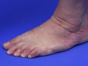
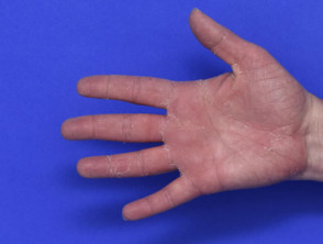
Keratolytic winter erythema usually presents between infancy and early adulthood, and it continues with an intermittent and recurrent pattern [2]. It ranges in severity with cyclical skin peeling and underlying erythema affecting the palms of the hands and the soles of the feet [3].
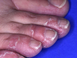
- A flare begins with the erythema. This may be annular and involve the webbed spaces of the fingers and toes or the entire palm or sole. The erythema can be transgradient, spreading to the top of the hands and feet.
- Erythema is followed by painless superficial dry blisters or opaque-appearing skin that peels centrifugally.
- Each patch expands over 4–6 weeks before healing [1].
- The erythema retains the papillary ridge pattern of the fingerprints.
- Palmoplantar sweating may lead to a pungent odour and increased peeling.
Some patients with keratolytic winter erythema also develop annular erythema on the limbs, buttocks, or trunk; there has been one reported case of facial involvement [4].
Aggravating factors may include:
- The winter season
- Excessive exposure to water, chemicals, or friction [2]
- Illness
- Psychological stress
- Menstruation
- Secondary bacterial infection
- Certain medications including topical steroids and general anaesthesia.
Keratolytic winter erythema can improve in summer and during pregnancy [3].
What are the complications of keratolytic winter erythema?
Active flares of keratolytic winter erythema can be disabling in severe cases.
How is keratolytic winter erythema diagnosed?
The diagnosis of keratolytic winter erythema may be evident clinically and may be supported by a positive family history.
On skin biopsy, keratolytic winter erythema shows characteristic histopathology with basal keratinocyte proliferation and defective layers of the stratum corneum [5].
What is the differential diagnosis for keratolytic winter erythema?
Depending on the phase of the lesion, the differential diagnosis of keratolytic winter erythema may include [6]:
- Keratolysis exfoliativa
- Palmoplantar keratoderma
- Localised epidermolysis bullosa simplex
- Circumscribed palmar hypokeratosis
- Annular erythema
- Erythrokeratoderma.
What is the treatment for keratolytic winter erythema?
There is currently no established effective treatment for keratolytic winter erythema. Topical keratolytics, retinoids and steroids may aggravate keratolytic winter erythema [6]. Photodynamic therapy has shown disease-modifying results in one patient [5].
What is the outcome for keratolytic winter erythema?
Keratolytic winter erythema typically improves or even clears during the summer months [4]. It also tends to improve with age and there may be only minimal scaling in the creases in adulthood [1]. In some severely affected individuals, it will persist [2].