Histology of Mycobacterium marinum
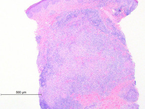
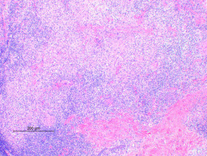
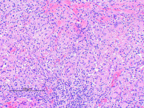
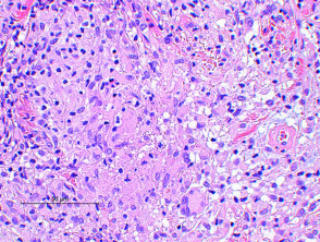
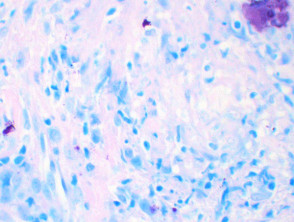
The histologic findings of Mycobacterium marinum infection vary by the age of the lesion. Scanning power view of well developed lesions demonstrate a granulomatous dermatitis (Figure 1), forming an extensive inflammatory nodular infiltrate within the dermis. Early lesions may show an acute suppurative inflammatory process with little granuloma formation. The epidermis may show prominent pseudoepitheliomatous hyperplasia with or without ulceration. There are tuberculoid granulomas with varying degrees of abscess formation (Figure 2). The infiltrate is mixed lymphohistiocytic with multinucleated giant cells and scattered neutrophils (Figures 3 and 4).
Mycobacterium marinum skin infection pathology
Special stains in Mycobacterium marinum skin infection
Acid fast stains such as Ziehl-Neelsen (Figure 5) reveal bacteria in the majority of cases, but can be negative. Culture at 25-30 Celsius is therefore essential to making a diagnosis in cases with suspicious histology.
Mycobacterium marinum skin infection – Ziehl-Neelsen stain
Differential diagnosis of Mycobacterium marinum skin infection
Atypical infections: Other mycobacterial organisms need to be considered based on histologic findings alone. Neutrophil infiltrates, interstitial granulomas, small vessel proliferation and high numbers of bacilli are more frequently seen in non-tuberculous (over tuberculous) mycobacterial infections. Cultures studies and PCR are essential for species identification. Deep fungal infections also show suppurative granuloma formation, with fungal elements visible on PAS or silver staining.
Interstitial granuloma annulare: Rare cases have been described where the histology showed an interstitial histiocytic infiltrate resembling interstitial granuloma annulare. Acid fast staining revealed bacterial organisms.