What is paederus dermatitis?
Paederus dermatitis is a skin irritation due to contact with certain species of the rove beetle, such as the Nairobi fly. It is also known as rove beetle rash, dermatitis linearis, spider lick, night burn, and Nairobi fly rash.
A blistering rash occurs 24–48 hours after brushing against or crushing the beetle against the skin, and can take several weeks to disappear [1,2].
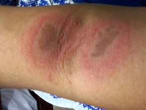
Paedarus dermatitis
Who gets paederus dermatitis?
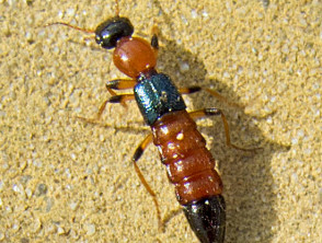
At least 60,000 different species of rove beetle have been identified — the largest group of insects worldwide. These insects are of the family Staphylinidae, in the order Coleoptera (beetles) [2,3]. The beetles have narrow bodies, ranging from 0.5–1.5 cm [4]. They tend to have a shiny black head and thorax, with blue or black elytra (forewing), and an orange-red abdomen [1,4].
Rove beetles can be found in decaying vegetable and animal matter in most environments around the world, except Antarctica [4]. There are more than 1000 different species of rove beetle in New Zealand. They are more prevalent in warmer climates [5]. The beetle breeding period is during the rainy seasons [1,4].
Paederus dermatitis is due to contact with one of more than 622 paederus species of rove beetle, which have a blistering agent in their haemolymph (haemolymph is analogous to blood in vertebrates).
Outbreaks of paederus dermatitis are most commonly reported in Europe and Asia, but outbreaks have occurred in many other countries including:
- Australia
- Malaysia
- Sri Lanka
- Kenya
- Iran
- Central Africa
- Uganda
- Okinawa
- Sierra Leone
- Argentina
- Brazil
- France
- Venezuela
- Ecuador
- India [2,5].
The beetle is attracted to ultraviolet radiation (UVR); epidemics have been reported in warm regions with military units and hospital wards with open windows and fluorescent lights [4].
Paederus beetle
What causes paederus dermatitis?
Paederus dermatitis is due to paederin, a toxin produced by pseudomonas bacteria in the haemolymph and released by the female paederus beetle [2,4,7]. Paederin causes a release of epidermal proteases and a loss of intercellular connection, inhibiting protein synthesis, DNA synthesis, and mitosis [2,5].
What are the clinical features of paederus dermatitis?
A localised streaky or linear erythema appears 24–48 hours after contact with the beetle and is typically followed by vesicles and pustules after 2–4 days [2,4,8]. Signs take a week or more to disappear [2].
The cutaneous features of paederus dermatitis include:
- Erythema [1,4]
- Vesicles and pustules [4,8]
- A burning sensation [1,4–8]
- ‘Kissing lesions’ where two adjacent flexural surfaces come together [1,4]
- Periocular dermatitis and keratoconjunctivitus (‘Nairobi eye’) [1,2,4,8]
- Balanitis (inflammation of the glans of the penis) due to transfer of the toxin on hands [1,2].
What are the complications of paederus dermatitis?
The primary complication of paederus dermatitis is the pain associated with the rash. Secondary complications include:
- Infection
- Exfoliation and ulceration (sometimes requiring hospitalisation)
- Postinflammatory hyperpigmentation
- Scarring [2,5].
How is paederus dermatitis diagnosed?
Paederus dermatitis is diagnosed clinically.
- A skin biopsy of an early lesion shows neutrophilic spongiosis, vesiculation, and reticular necrosis of the epidermis [4]. The inflammatory infiltrate in the epidermis contains many neutrophils [4,8].
- Later biopsies show irregular acanthosis, pallor of superficial keratinocytes, overlying parakeratosis, confluent epidermal necrosis, and suprabasal acantholysis [7,8].
What is the differential diagnosis for paederus dermatitis?
Paederus dermatitis may be confused with:
- Herpes simplex
- Herpes zoster
- Acute allergic contact dermatitis or irritant contact dermatitis
- Phytophotodermatitis
- Bullous impetigo
- Scald
- Chemical burn
- Cutaneous larva migrans
- Dermatitis herpetiformis
- Pemphigus foliaceus
- Caterpillar dermatitis
- Millipede dermatitis
- Moth-related dermatitis
- Meloid beetle toxin-induced vesicular dermatitis
- Lax beetle dermatosis
- Dermatitis artefacta
- Trichinosis [2,5,6].
The distinguishing features of paederus dermatitis include [1]:
- Irritation confined to exposed areas
- Kissing lesions
- Occurrence during rainy/warm season
- Other individuals presenting with similar lesions
- Histopathology.
What is the treatment for paederus dermatitis?
Once symptoms have appeared, the initial step should be to wash the affected area with soap and clean water to remove the pederin toxin.
After cleaning the area, apply a cold wet compress and a topical steroid [1].
- Tincture of iodine can help neutralise the paederin toxin and act as an antiseptic [5].
- Soothing creams containing calamine, camphor, and topical anaesthetic can provide relief of pain and itching [5].
- Oral antibiotics (eg, ciprofloxacin) for secondary bacterial infection [7].
How can paederus dermatitis be prevented?
Paederus dermatitis can be prevented by limiting the chance of exposure to the rove beetle.
- Use insect-proof netting at night.
- Select light sources that do not emit UV.
- Turn lights off when sleeping.
- Remove any beetle found on the skin without crushing it.
- Wash skin in contact with a rove beetle with soap and water [4,5,7].
What is the outcome for paederus dermatitis?
It can take a few weeks for paederus dermatitis to resolve, and postinflammatory pigmentation may persist for several months [1,2].