What is pilonidal disease?
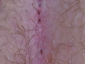
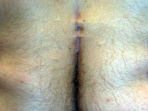
Pilonidal disease is a chronic skin problem found most often in the sacrococcygeal region. This is the cleft between the buttocks just below the base of the spine. It is characterised by one or more sinus tracts; these are cavities with a narrow opening on the skin surface (pilonidal sinus). In most cases, the cavity is filled with nests of hair – hence the name pilonidal ("pilus" meaning hair and "nidal" meaning nest). A non-inflamed lump is known as a pilonidal cyst. If the sinus becomes infected a pilonidal abscess may form.
Pilonidal sinuses
What causes pilonidal disease?
The exact reason why pilonidal disease occurs is still unclear. Possible causes include:
- Some people are born with small holes or pits near the base of the spine. These are in fact enlarged hair follicles.
- Follicular occlusion; some people are genetically prone to this. They may also suffer from hidradenitis suppurativa, acne conglobata, and dissecting cellulitis (follicular occlusion syndrome or tetrad).
- When subjected to friction and motion, the follicles are injured and disrupted so the hair pokes through the wall of the follicle into the surrounding skin setting up a foreign body reaction.
- Neighbouring hairs or free hairs from other parts of the body collect in the pit and invade the small opening created by the distorted hair follicles.
- Skin and perineal bacteria such as Staphylococcus aureus and Bacteroides species invade the opening and cause infection.
Who gets pilonidal disease?
Pilonidal disease affects both men and women usually between the ages of 20–40 years. It is 2–3 times more common in men than women. Other factors that increase the risk of pilonidal disease include:
- Coarse, curly or crinkly hair
- Obesity
- Family predisposition
- Poor hygiene
- Prolonged sitting or buttock friction causing increased sweating
- Repeated local injury (once known as "Jeep rider's disease" as it hospitalised more than 80,000 US soldiers in WWII)
- Co-existing hidradenitis suppurativa
What are the signs and symptoms?
Signs and symptoms can vary from a small painless pit or dimple at the base of the spine to a large painful abscess. Most patients have progressive tenderness, particularly after prolonged periods of sitting, such as during a long drive. Signs and symptoms include:
- Pain, redness and swelling
- Small hole or holes draining fluid that may be clear, cloudy or bloody
- If infected, the draining pus may have a foul odour
- Fever, malaise or nausea
- Visible or lumpy tracts 2–5 cm long in chronic or recurrent pilonidal disease
How is the diagnosis made?
The clinical features of pilonidal sinus is usually straightforward. If necessary, skin biopsy can be undertaken. The histopathological features of pilonidal sinus characteristically show foreign body reaction.
What treatment is available?
A pilonidal cyst that isn't causing any problems doesn't require any treatment. The patient should be advised to keep the area clean and free of hair by shaving or using a hair removal agent every 2–3 weeks. The cyst may resolve itself. Persistent and inflamed cysts (acute pilonidal abscess) are incised (cut into) and drained out to reduce inflammation and pain. Occasionally the abscess cavity may be cut out completely to remove hair nests and skin debris; this reduces the rate of recurrence to about 15%.
Persistent, complex or recurrent pilonidal sinus disease must be treated surgically. Procedures vary from taking the roof off the sinuses to wide and deep excision (ie all affected areas are completely cut out). In all cases, the cavity is scrubbed and scraped out to remove hair and abnormally healing granulation tissue. Several techniques are available for wound healing and closure; these include
- Dressing or packing open wounds
- Marsupialisation (forming a pouch), which results in a smaller wound compared to wounds that are left open to granulate
- Closure using skin flaps for wide excisions.