DermNet provides Google Translate, a free machine translation service. Note that this may not provide an exact translation in all languages

Translate

Radiographic investigations in melanoma

Author: Dr Matthew Howard, Clinic/Research Fellow, Victorian Melanoma Service, Melbourne, VIC. Australia. Reviewed by Dr Martin Cherk, Nuclear Medicine Physician and Medical Oncologist, Alfred Health, Melbourne, VIC, Australia. DermNet NZ Editor in Chief: Adjunct A/ Prof. Amanda Oakley, Dermatologist, Hamilton, New Zealand. Copy edited by Gus Mitchell. January 2020.


toc-icon

What are radiographic investigations?

Radiographic investigations produce images of tissues and bones under the skin. The term is often used to include a variety of imaging methods using X-rays, computed tomography (CT) scans, ultrasound scans, and magnetic resonance imaging (MRI).

Radiographic investigations in melanoma

Why are radiographic investigations in melanoma important?

There are benefits for melanoma patients from radiographic investigations, such as the early detection of metastases or for reassurance, but there are also risks, such as detection of unimportant lesions (false-positives) or missed metastases (false negatives). Follow-up should include evaluation of any symptoms and examination of the site of the original tumour and regional lymph nodes. Regular skin examinations are also important as radiographic investigations rarely detect new primary melanomas.

There are three key indications for radiographic investigations of patients with melanoma. 

  • Initial staging of a patient with melanoma to determine if high-risk cancer has spread to any internal body sites
  • To evaluate the response to treatment (eg, targeted therapies and immunotherapies) [1–3]
  • To monitor patients after melanoma resection for patients at high risk of recurrence or progression with the aim of detecting early recurrence [1].

Guidelines and standards indicate that investigations are not indicated for melanoma in situ (Stage 0) or thin invasive melanoma (Stage I or II) with no clinical symptoms or signs of metastasis. The appropriate patients for staging or surveillance are those at significant risk for distant metastases [4].

Although primary melanoma tumours and local metastases in the lymph nodes or in-transit can often be detected on clinical examination, clinical monitoring for deep lymph nodes and distant viscera is more difficult. Distant visceral metastatic disease is often asymptomatic until advanced, when it may be difficult to remove surgically [1].

What radiographic investigations are used in the staging and surveillance of malignant melanoma?

Basic radiological investigations such as chest x-ray (CXR) can detect occult metastatic melanoma, but their two-dimensional soft-tissue views are limited [5]. Thus many patients will be referred for CT, positron emission tomography-CT (PET-CT), ultrasound imaging, and MRI scans.

Computed tomography and positron emission tomography-CT

CT and PET-CT scans are commonly used in staging and surveillance of many solid malignancies.

  • CT provides three-dimensional views of the body.
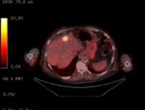
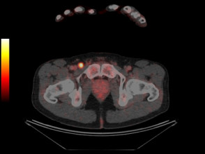
  • PET-CT adds a fluorodeoxyglucose (FDG) tracer, which highlights areas of increased metabolically activity frequently seen with solid malignancies including melanoma.
  • PET-CT provides incremental information to CT alone as it can detect metabolically active disease in tissues such as lymph nodes which may appear structurally normal on CT.
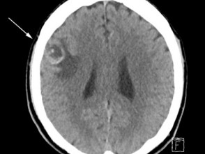
  • PET-CT can accurately detect metastases in most body sites, except the brain where there is high background metabolic activity and diagnostic CT or MRI is superior [6].

Ultrasound imaging

Ultrasound imaging is used to monitor regional lymph node basins for metastases. The accuracy of ultrasound is dependent upon the skill and experience of the sonographer [7,8].

Magnetic resonance imaging

MRI provides high-resolution imaging.

  • It is used to look for metastatic disease in the brain.
  • MRI may be also used to investigate suspicious areas seen on PET-CT prior to surgery or biopsy [9].

Which patients should undergo radiographic investigations for melanoma surveillance?

Radiographic investigations are appropriate for patients with stage III metastatic melanoma, where there has been histologically confirmed spread of melanoma to lymph nodes, in-transit metastases, or satellite metastases. These patients are at high risk of recurrence or progression to stage IV metastatic melanoma [10–12]. The relative poor mortality of stage IIC melanoma also merits consideration of surveillance [12].

  • International guidelines for surveillance imaging in melanoma patients are inconsistent as they are based upon expert consensus rather than evidence.
  • The results of published research studies are difficult to compare because they have enrolled patients with melanoma at differing stages.
  • Until recently, there were no effective systemic treatment options, so there was little interest in evaluating radiographic investigations. This has changed with the development of targeted therapies and immunotherapies.
  • The American National Comprehensive Cancer Network (NCCN) recommends CT or CT-PET scans 3–12 monthly for patients with asymptomatic stage IIB–IV melanoma [13].
  • The European Society of Medical Oncology recommends only physical examination every three months [14].

Ultrasound surveillance of the regional nodal basin can be considered for:

  • Surveillance for recurrence in patients who have a positive sentinel lymph node biopsy and who have not undergone complete lymph node dissection.
  • Surveillance in those who have not had a sentinel lymph node biopsy but remain at high risk of progression to stage III disease due to unfavourable characteristics of the primary tumour.
  • Initial staging of the lymph node basin; however, ultrasound is not as accurate as sentinel lymph node biopsy [15–17].

Ultrasound surveillance in stage I or II melanoma is superior to the clinical examination of the regional lymph node, but the long-term survival benefit of ultrasound surveillance is unknown [8]. Only a few patients benefit from earlier detection of lymph node metastasis or avoidance of unnecessary surgery, and some undergo unnecessary surgery for suspicious findings [8].

What are the benefits of radiographic investigations for melanoma surveillance?

Isolated metastases detected by surveillance or staging imaging can be potentially cured by surgical resection, radiotherapy, or systemic therapies [18–20], and patients have superior survival rates with modern therapies [21–23].

  • In Australian data, PET detected metastases up to six months earlier than other imaging or physical examinations [24].
  • Two studies showed a patient’s treatment changed in 19–35% of stage III melanoma patients following PET-CT scans [25–27]
  • PET-CT and CT are also able to detect additional primary malignancies [28–29]. 

Other advantages of selected imaging modalities

  • Ultrasound imaging is non-invasive and does not emit radiation.
  • MRI does not emit radiation, unlike CT scans.

What are the controversies and disadvantages of radiographic surveillance of melanoma?

Without a randomised trial, it is difficult to prove that melanoma survival is superior in those who have undergone imaging.

Even after negative imaging, patients or their partners detect the majority of recurrences in melanoma as they are often visible or palpable within the skin or lymph nodes [30,31].

  • The detection rates for imaging of asymptomatic distant metastases vary between 15 and 72% depending on staging [31–33].
  • PET-CT rarely detects metastatic disease smaller than 5 mm [34–36].
  • The benefit of PET-CT in detecting occult metastases in sentinel lymph node-positive patients ranged from 0.5 to 3.7% [37–40].

There is a risk that surveillance imaging may detect non-specific lesions or result in false-positive findings for metastasis [1,41].

  • Additional invasive investigations may be undertaken when there are non-specific findings or false positives [41,42].
  • PET surveillance in stage IIIA metastatic melanoma patients has been associated with a 7–14% false-positive rate [1,43,44].
    • In one of these studies, 86% of these false-positive findings underwent biopsy [1].

Multiple studies have evaluated the overall accuracy of PET-CT staging and surveillance.

  • Sensitivity and specificity of PET-CT was 65% and 99% for regional disease,  86% and 91% for distant metastasis, and 80% and 87% for staging respectively [45].
  • Ultrasonography had the highest sensitivity (60%) and specificity (97%) for the staging of regional lymph nodes and 96% and 99% for targeted lymph node respectively [45].
  • The reliability of PET and CT brain in detecting brain metastasis is poor [46,47]. In one study of almost 700 patients with cutaneous metastatic melanoma, 12% were found to have asymptomatic brain metastases on CT scan [48].
  • MRI brain is superior for evaluation of intracranial metastatic disease but is contraindicated in patients with metal implants.
  • A single PET-CT detected 24% of recurrences in asymptomatic melanoma in 110 asymptomatic patients with stage IIB to IIIB melanoma [44].
  • In 170 patients undergoing PET-CT surveillance imaging, melanoma recurrence was identified in 38%, of which 69% were asymptomatic. A negative PET-CT at 18 months had negative predictive values 80–84% for true non-recurrence at any time in the 47-month (median) follow-up period [1]. Of the PET-CT detected recurrent patients, 33 (52%) underwent potentially curative resection, although only 16% remained disease-free after 24 months [1].

How often should surveillance scans be performed?

The optimum frequency for surveillance scans — and for how long — are unknown.

  • Typically, PET scans are performed every 6 months for surveillance of high-risk patients [1,23].
  • Most recurrences of melanoma occur within the first two to three years [1,11].


References

  1. Lewin J, Sayers L, Kee D, et al. Surveillance imaging with FDG-PET/CT in the post-operative follow-up of stage 3 melanoma. Ann Oncol. 2018 Jul 1;29(7):1569–74. doi: 10.1093/annonc/mdy124. PMID: 29659679. PubMed
  2. Arrangoiz R, Papavasiliou P, Stransky CA, et al. Preoperative FDG-PET/CT is an important tool in the management of patients with thick (T4) melanoma. Dermatol Res Pract. 2012;2012:614349. doi: 10.1155/2012/614349. Epub 2012 May 13. PMID: 22654898; PMCID: PMC3359680. PubMed
  3. Cho S, Lipson E, Im H, et al. Prediction of response to immune checkpoint inhibitor therapy using early-time-point 18F-FDG PET/CT imaging in patients with advanced melanoma. J Nucl Med. 2017 Sep;58(9):1421–8. doi: 10.2967/jnumed.116.188839. Epub 2017 Mar 30. PMID: 28360208; PMCID: PMC5577627. PubMed
  4. Fink A, Holle-Robatsch S, Herzog N, et al. Positron emission tomography is not useful in detecting metastasis in the sentinel lymph node in patients with primary malignant melanoma stage I and II. Melanoma Res. 2004 Apr;14(2):141–5. doi: 10.1097/00008390-200404000-00011. PMID: 15057045. PubMed
  5. Terhune M, Swanson N, Johnson T. Use of chest radiography in the initial evaluation of patients with localized melanoma. Arch Dermatol. 1998 May;134(5):569–72. doi: 10.1001/archderm.134.5.569. PMID: 9606326. PubMed
  6. Schröer-Günther MA, Wolff RF, Westwood ME, et al. F-18-fluoro-2-deoxyglucose positron emission tomography (PET) and PET/computed tomography imaging in primary staging of patients with malignant melanoma: a systematic review. Syst Rev. 2012 Dec 13;1:62. doi: 10.1186/2046-4053-1-62. PMID: 23237499; PMCID: PMC3536719. PubMed Central
  7. Bafounta M, Beauchet A, Chagnon S, Saiag P. Ultrasonography or palpation for detection of melanoma nodal invasion: a meta-analysis. Lancet Oncol. 2004 Nov;5(11):673-80. doi: 10.1016/S1470-2045(04)01609-2. PMID: 15522655. PubMed
  8. Machet L, Nemeth-Normand F, Giraudeau B, et al. Is ultrasound lymph node examination superior to clinical examination in melanoma follow-up? A monocentre cohort study of 373 patients. Br J Dermatol. 2005 Jan;152(1):66–70. doi: 10.1111/j.1365-2133.2004.06262.x. PMID: 15656802. PubMed
  9. Deike-Hofmann K, Thünemann D, Breckwoldt M, et al. Sensitivity of different MRI sequences in the early detection of melanoma brain metastases. PLoS One. 2018 Mar 29;13(3):e0193946. doi: 10.1371/journal.pone.0193946. PMID: 29596475; PMCID: PMC5875773. PubMed
  10. Leiter U, Meier F, Schittek B, Garbe C. The natural course of cutaneous melanoma. J Surg Oncol. 2004 Jul 1;86(4):172–8. doi: 10.1002/jso.20079. PMID: 15221923. PubMed
  11. Romano E, Scordo M, Dusza S, Coit D, Chapman P. Site and timing of first relapse in stage III melanoma patients: implications for follow-up guidelines. J Clin Oncol. 2010 Jun 20;28(18):3042–7. doi: 10.1200/JCO.2009.26.2063. Epub 2010 May 17. PMID: 20479405; PMCID: PMC3664035. PubMed
  12. Gershenwald JE, Scolyer RA, Hess KR, et al. Melanoma staging: evidence-based changes in the American Joint Committee on Cancer eighth edition cancer staging manual. CA Cancer J Clin. 2017;67(6):472–492. doi:10.3322/caac.21409 PubMed
  13. Coit D, Andtbacka R, Bichakjian C,  et al. Melanoma. J Natl Compr Canc Netw. 2009 Mar;7(3):250–75. doi: 10.6004/jnccn.2009.0020. PMID: 19401060.PubMed
  14. Dummer R, Hauschild A, Pentheroudakis G. Cutaneous malignant melanoma: ESMO clinical recommendations for diagnosis, treatment and follow-up. Ann Oncol. 2009 May;20 Suppl 4:129–31. doi: 10.1093/annonc/mdp152. PMID: 19454433. PubMed
  15. Chai C, Zager J, Szabunio M, et al. Preoperative ultrasound is not useful for identifying nodal metastasis in melanoma patients undergoing sentinel node biopsy: preoperative ultrasound in clinically node-negative melanoma. Ann Surg Oncol. 2012 Apr;19(4):1100–6. doi: 10.1245/s10434-011-2172-7. Epub 2011 Dec 23. PMID: 22193886; PMCID: PMC4504035. PubMed
  16. Ogata D, Uematsu T, Yoshikawa S, Kiyohara Y. Accuracy of real-time ultrasound elastography in the differential diagnosis of lymph nodes in cutaneous malignant melanoma (CMM): a pilot study. Int J Clin Oncol. 2014 Aug;19(4):716–21. doi: 10.1007/s10147-013-0595-9. Epub 2013 Jul 31. PMID: 23900625. PubMed
  17. Stoffels I, Dissemond J, Poeppel T, et al. Advantages of preoperative ultrasound in conjunction with lymphoscintigraphy in detecting malignant melanoma metastases in sentinel lymph nodes: a retrospective analysis in 221 patients with malignant melanoma AJCC Stages I and II. J Eur Acad Dermatol Venereol. 2012 Jan;26(1):79–85. doi: 10.1111/j.1468-3083.2011.04013.x. Epub 2011 Mar 14. PMID: 21395693. PubMed
  18. Sosman J, Moon J, Tuthill R, et al. A phase 2 trial of complete resection for stage IV melanoma: results of Southwest Oncology Group Clinical Trial S9430. Cancer. 2011 Oct 15;117(20):4740–06. doi: 10.1002/cncr.26111. Epub 2011 Mar 31. PMID: 21455999; PMCID: PMC3129414. PubMed
  19. Wasif N, Bagaria S, Ray P, Morton D. Does metastasectomy improve survival in patients with Stage IV melanoma? A cancer registry analysis of outcomes. J Surg Oncol. 2011 Aug 1;104(2):111–5. doi: 10.1002/jso.21903. Epub 2011 Mar 4. PMID: 21381040; PMCID: PMC3199373. PubMed
  20. He M, Lovell J, Ng B, Spillane J, et al. Post-operative survival following metastasectomy for patients receiving BRAF inhibitor therapy is associated with duration of pre-operative treatment and elective indication. J Surg Oncol. 2015 Jun;111(8):980–4. doi: 10.1002/jso.23938. Epub 2015 Jun 17. PMID: 26080731. PubMed
  21. Long G, Weber J, Infante J, et al. Overall survival and durable responses in patients with BRAF V600-mutant metastatic melanoma receiving dabrafenib combined with trametinib. J Clin Oncol. 2016 Mar 10;34(8):871–8. doi: 10.1200/JCO.2015.62.9345. Epub 2016 Jan 25. Erratum in: J Clin Oncol. 2019 Feb 1;37(4):355. PMID: 26811525. PubMed
  22. Joseph RW, Elassaiss-Schaap J, Wolchok JD, et al (2014). Baseline tumor size as an independent prognostic factor for overall survival in patients with metastatic melanoma treated with the anti-PD-1 monoclonal antibody MK-3475. J Clin Oncol. 2014;32(15): 3015. https://doi.org/10.1200/jco.2014.32.15_suppl.3015.
  23. Lim KHJ, Spain L, Barker C, et al. Contemporary outcomes from the use of regular imaging to detect relapse in high-risk cutaneous melanoma. ESMO Open. 2018 Feb 24;3(2):e000317. doi: 10.1136/esmoopen-2017-000317. PMID: 29531842; PMCID: PMC5844377. PubMed
  24. Damian D, Fulham M, Thompson E, Thompson J. Positron emission tomography in the detection and management of metastatic melanoma. Melanoma Res. 1996 Aug;6(4):325–9. doi: 10.1097/00008390-199608000-00008. PMID: 8873053. PubMed
  25. Davis PC, Hudgins PA, Peterman SB, Hoffman JC Jr. Diagnosis of cerebral metastases: double-dose delayed CT vs contrast-enhanced MR imaging. AJNR Am J Neuroradiol. 1991 Mar-Apr;12(2):293–300. PMID: 1902031. PubMed
  26. Bastiaannet E, Wobbes T, Hoekstra O, et al. Prospective comparison of [18F]fluorodeoxyglucose positron emission tomography and computed tomography in patients with melanoma with palpable lymph node metastases: diagnostic accuracy and impact on treatment. J Clin Oncol. 2009 Oct 1;27(28):4774–80. doi: 10.1200/JCO.2008.20.1822. Epub 2009 Aug 31. PMID: 19720925. PubMed
  27. Brady M, Akhurst T, Spanknebel K, et al.  Utility of preoperative [(18)]f fluorodeoxyglucose-positron emission tomography scanning in high-risk melanoma patients. Ann Surg Oncol. 2006 Apr;13(4):525-32. doi: 10.1245/ASO.2006.02.008. Epub 2006 Feb 15. PMID: 16474909.PubMed
  28. Ishimori T, Patel P, Wahl R. Detection of unexpected additional primary malignancies with PET/CT. J Nucl Med. 2005 May;46(5):752–7. PMID: 15872346. PubMed
  29. Lumbreras B, Donat L, Hernández-Aguado I. Incidental findings in imaging diagnostic tests: a systematic review. Br J Radiol. 2010 Apr;83(988):276–89. doi: 10.1259/bjr/98067945. PMID: 20335439; PMCID: PMC3473456. PubMed
  30. Meyers M, Yeh J, Frank J, et al. Method of detection of initial recurrence of stage II/III cutaneous melanoma: analysis of the utility of follow-up staging. Ann Surg Oncol. 2009 Apr;16(4):941–7. doi: 10.1245/s10434-008-0238-y. Epub 2008 Dec 20. PMID: 19101766. PubMed
  31. Garbe C, Paul A, Kohler-Späth H, et al. Prospective evaluation of a follow-up schedule in cutaneous melanoma patients: recommendations for an effective follow-up strategy. J Clin Oncol. 2003 Feb 1;21(3):520–9. doi: 10.1200/JCO.2003.01.091. PMID: 12560444. PubMed
  32. Hofmann U, Szedlak M, Rittgen W, Jung EG, Schadendorf D. Primary staging and follow-up in melanoma patients--monocenter evaluation of methods, costs and patient survival. Br J Cancer. 2002 Jul 15;87(2):151–7. doi: 10.1038/sj.bjc.6600428. PMID: 12107834; PMCID: PMC2376106. PubMed
  33. Hengge UR, Wallerand A, Stutzki A, Kockel N. Cost-effectiveness of reduced follow-up in malignant melanoma. J Dtsch Dermatol Ges. 2007 Oct;5(10):898-907. English, German. doi: 10.1111/j.1610-0387.2007.06454.x. PMID: 17910672. PubMed
  34. Akcali C, Zincirkeser S, Erbagcý Z, et al. Detection of metastases in patients with cutaneous melanoma using FDG-PET/CT. J Int Med Res. 2007 Jul-Aug;35(4):547–53. doi: 10.1177/147323000703500415. PMID: 17697533. PubMed
  35. Strobel K, Dummer R, Husarik DB, Pérez Lago M, Hany TF, Steinert HC. High-risk melanoma: accuracy of FDG PET/CT with added CT morphologic information for detection of metastases. Radiology. 2007 Aug;244(2):566-74. doi: 10.1148/radiol.2442061099. PMID: 17641374. PubMed
  36. Stas M, Stroobants S, Dupont P, et al. 18-FDG PET scan in the staging of recurrent melanoma: additional value and therapeutic impact. Melanoma Res. 2002 Oct;12(5):479–90. doi: 10.1097/00008390-200209000-00010. PMID: 12394190. PubMed
  37. Aloia T, Gershenwald J, Andtbacka R, et al. Utility of computed tomography and magnetic resonance imaging staging before completion lymphadenectomy in patients with sentinel lymph node-positive melanoma. J Clin Oncol. 2006 Jun 20;24(18):2858–65. doi: 10.1200/JCO.2006.05.6176. PMID: 16782925. PubMed
  38. Gold J, Jaques D, Busam K, Brady M, Coit D. Yield and predictors of radiologic studies for identifying distant metastases in melanoma patients with a positive sentinel lymph node biopsy. Ann Surg Oncol. 2007 Jul;14(7):2133–40. doi: 10.1245/s10434-007-9399-3. Epub 2007 Apr 24. PMID: 17453294. PubMed
  39. Miranda E, Gertner M, Wall J, et al.Routine imaging of asymptomatic melanoma patients with metastasis to sentinel lymph nodes rarely identifies systemic disease. Arch Surg. 2004 Aug;139(8):831–6; discussion 836–7. doi: 10.1001/archsurg.139.8.831. PMID: 15302691. PubMed
  40. Pandalai P, Dominguez F, Michaelson J, Tanabe K. Clinical value of radiographic staging in patients diagnosed with AJCC stage III melanoma. Ann Surg Oncol. 2011 Feb;18(2):506–13. doi: 10.1245/s10434-010-1272-0. Epub 2010 Aug 24. PMID: 20734149. PubMed
  41. Leon-Ferre R, Kottschade L, Block M, et al. Association between the use of surveillance PET/CT and the detection of potentially salvageable occult recurrences among patients with resected high-risk melanoma. Melanoma Res. 2017 Aug;27(4):335–41. doi: 10.1097/CMR.0000000000000344. PMID: 28296712. PubMed
  42. Warner E. Intensive radiologic surveillance: a focus on the psychological issues. Ann Oncol. 2004;15 Suppl 1:I43-7. doi: 10.1093/annonc/mdh657. PMID: 15280187. PubMed
  43. Baker J, Meyers M, Frank J, Amos K, Stitzenberg K, Ollila D.Routine restaging PET/CT and detection of initial recurrence in sentinel lymph node positive stage III melanoma. Am J Surg. 2014 Apr;207(4):549–54. doi: 10.1016/j.amjsurg.2013.04.012. PMID: 24674829. PubMed
  44. Koskivuo I, Kemppainen J, Giordano S, et al. Whole body PET/CT in the follow-up of asymptomatic patients with stage IIB-IIIB cutaneous melanoma. Acta Oncol. 2016 Nov;55(11):1355–9. doi: 10.1080/0284186X.2016.1213879. Epub 2016 Aug 24. PMID: 27553064. PubMed
  45. Xing Y, Bronstein Y, Ross M, et al. Contemporary diagnostic imaging modalities for the staging and surveillance of melanoma patients: a meta-analysis. J Natl Cancer Inst. 2011 Jan 19;103(2):129–42. doi: 10.1093/jnci/djq455. Epub 2010 Nov 16. PMID: 21081714; PMCID: PMC3022618. PubMed
  46. Bochev P, Klisarova A, Kaprelyan A, Chaushev B, Dancheva Z. Brain metastases detectability of routine whole body (18)F-FDG PET and low dose CT scanning in 2502 asymptomatic patients with solid extracranial tumors. Hell J Nucl Med. 2012 May-Aug;15(2):125–9. doi: 10.1967/s002449910030. Epub 2012 Jun 27. PMID: 22741148. PubMed
  47. Kitajima K, Nakamoto Y, Okizuka H, et al. Accuracy of whole-body FDG-PET/CT for detecting brain metastases from non-central nervous system tumors. Ann Nucl Med. 2008 Aug;22(7):595–602. doi: 10.1007/s12149-008-0145-0. Epub 2008 Aug 29. PMID: 18756362. PubMed
  48. Zukauskaite R, Schmidt H, Asmussen J, Hansen O, Bastholt L. Asymptomatic brain metastases in patients with cutaneous metastatic malignant melanoma. Melanoma Res. 2013 Feb;23(1):21–6. doi: 10.1097/CMR.0b013e32835ae915. PMID: 23117880. PubMed

On DermNet

Other websites

Books about skin diseases


Related information

Sign up to the newsletter